Ask a student who has only read about microscopy what a cell looks like, and they will give you a diagram from a textbook. Ask a student who has actually found one under a scope, and they will tell you about the moment the blur resolved into a wall, a nucleus, a chloroplast drifting past — the nudge of the fine focus, the light they had to get just right. The first student has a picture someone else drew. The second has a sighting — and the sighting is what the diagram was always trying to stand in for.
That gap is the whole reason this course is built the way it is. Microscopy is not a body of facts; it is a set of techniques. The entire living world it studies sits below the threshold of naked sight, and the only door into it is an instrument you have to learn to operate — carry, focus, mount, stain, resolve. The danger is that the subject collapses into staring at other people's photographs: a student learns to label a diagram of a cell without ever once bringing a real one into focus themselves.
The scope makes the invisible visible
The job of the microscope is to drag a whole hidden world up into view — but only for the hands that know how to drive it. You cannot see a plant cell, but you can peel a sliver of onion skin, mount it in a drop of water, lower a coverslip without trapping a bubble, and watch a tidy brick-wall of cells swim into focus under your own eye. You cannot see a single-celled organism unaided, but an instructor-approved specimen or recorded sequence can reveal its form and motion. Do not collect or culture unknown environmental or human material. You cannot grasp the difference between magnification and resolution by reading about it — but you can rack up to the high-power objective and discover, first-hand, that a bigger blur is still a blur.
This is what we mean when we say the course is skill-led, not textbook-led. The reading does not come first, with the scope as a garnish to confirm it. The bench comes first. The technique is performed where it actually lives (slide in hand, eye at the eyepiece), and the textbook is the tool we reach for to name what we just saw. A student who has watched cytoplasm stream inside a living leaf is ready to be told what they were looking at. A student who has only been told about it is ready to forget it.
The diagram in the book is a claim about something real. The scope is where the student finds out the claim is true — by seeing it themselves.
What the bench teaches that the page cannot
Beyond making the invisible concrete, the bench teaches a set of things a textbook structurally cannot, because they are not facts — they are techniques and judgments that only form under real conditions:
- That the instrument fights back. Finding focus without driving the objective into the slide, chasing a specimen that keeps drifting out of the field, coaxing enough light through a thick mount — these teach a patience with the tool that no worked example ever will.
- That real specimens don't look like the diagram. The cell is torn. The stain took unevenly. The structure you were sent to find is hiding behind a fold in the tissue. Learning to reason about why the real slide departs from the clean textbook drawing is a microscopist's core skill.
- That technique is knowledge. How you mount, how much stain, how you rack the focus, how you center and light the field — the result depends on the doing, and the doing can only be learned by doing.
The two-day rhythm
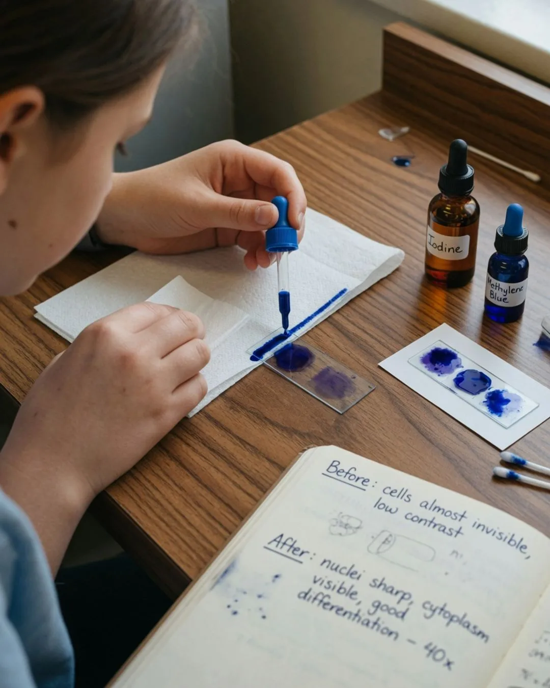
Practically, this conviction becomes a schedule. The course runs on a two-day rhythm. One day is the Concept Day: the technique is introduced and demonstrated up close — how a wet mount is built, how a stain adds contrast, how a scale bar is calibrated. The next is the Experiment Day, where that same technique becomes physical at the bench and gets written into a real lab notebook in the student's own hand. Between the two days, the student practices at home, and that gap is not dead time. It is where the technique settles into muscle memory.
We are not against the textbook; a serious microscopy course needs a rigorous one, and this course has it. We are against the textbook going first and the bench going second, because we have watched what that produces: a student who can recite the parts of a microscope and has never once brought a living cell into focus with their own hands. Put the bench first, and microscopy stops being a labeled diagram. It becomes a thing the student has actually seen, which is the only kind of microscopy anyone remembers.