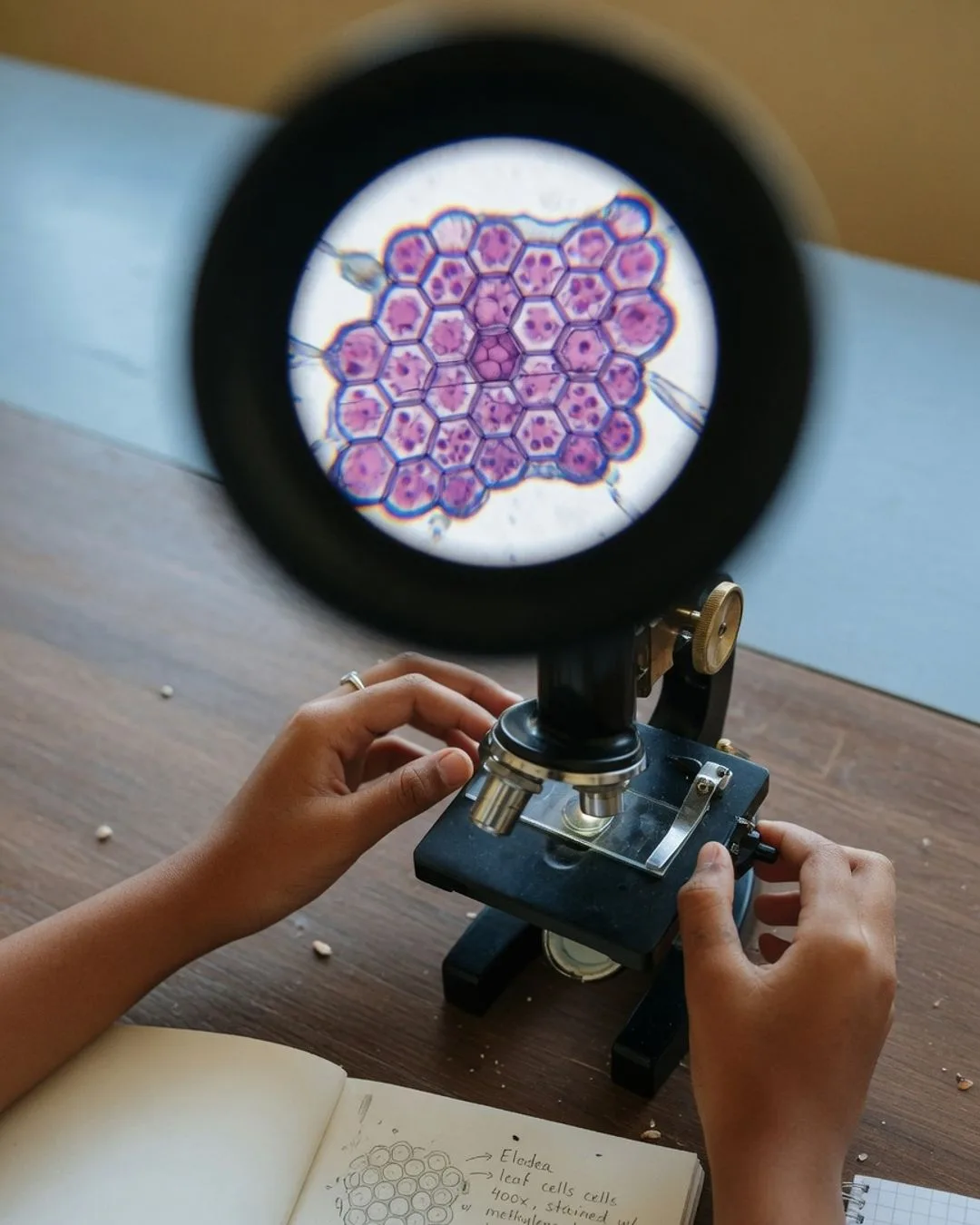
Every Bright Minds course has one unit where the walls between subjects come down on purpose — where microscopy refuses to stay in the microscopy box and pulls in history, reading, writing, and measurement because it cannot be honestly told without them. In this course, that unit is built around the founding of the science itself: Robert Hooke's Micrographia and Antonie van Leeuwenhoek's animalcules. It is the microscopy analog of the cholera map that anchors our biology course — a pair of real moments that turn out to touch everything.
The microscopy first
The instruments came first, and they were crude. In 1665 Robert Hooke (curator of experiments for the young Royal Society) pointed a compound microscope at a shaving of cork and saw it was not solid at all but a honeycomb of tiny empty chambers. He called them cells, after the bare rooms of a monastery, and the word stuck. His book, Micrographia, laid a fly's eye and a flea and the edge of a razor before the public in enormous, astonishing engravings, and it became the first scientific bestseller.
What makes this the perfect capstone is that seeing it at all required nearly everything the course teaches at once. It is a problem in magnification and resolution — Hooke and Leeuwenhoek were pushing single lenses to the ragged edge of what glass could do. It is a problem in specimen preparation: a fly's eye or a droplet of pond water has to be mounted and lit before it will give up its detail. It is a problem in observation and drawing — what you cannot capture on paper, you cannot share or defend. And it is a problem in measurement: Leeuwenhoek, a draper who ground his own lenses in secret, estimated the size of his "little animals" by comparing them to a grain of sand, and got astonishingly close. He did not get to master one skill; he had to hold all of them at once.
The same care a student brings to mounting a slide and drawing what they see is the care that, three hundred years ago, opened an entire kingdom of life no one had known was there.
The history and the Royal Society
Hooke and Leeuwenhoek worked at the height of the Scientific Revolution, in the first decades of the Royal Society — a moment when Europe decided that knowledge should come from looking rather than from authority. Hooke was at the Society's center, a paid experimenter and rival to Newton. Leeuwenhoek was an outsider: a cloth merchant in Delft with no Latin and no university, who ground lenses better than anyone alive and mailed his observations to London as a stream of plain-spoken letters. That the Society took a tradesman's word seriously — eventually electing him a Fellow — is itself a piece of history worth sitting with. It is the moment a new way of knowing began to outrank an old one, and it happened because two men could show, in a drawing and a letter, exactly what they had seen.
The reading and the writing
And then the course refuses to leave it at the pictures, because the discovery only counted once it was written down and read. Micrographia was as much a feat of prose and engraving as of optics — Hooke had to describe, in careful English, structures no reader had ever seen, and pair the words with plates good enough to be believed. Leeuwenhoek, writing in Dutch to a foreign society, had to make a skeptical audience trust a claim that sounded mad: that a single drop of rainwater teemed with thousands of living creatures too small to see. His letters are a masterclass in observational writing — precise, patient, insistent on detail.
We put those texts in front of students deliberately, because they teach something no diagram can:
- A discovery is not finished until it is communicated. Hooke and Leeuwenhoek saw wonders, but the science began only when they made others see them too — in words and drawings a stranger could trust. What you cannot describe, you have not fully understood.
- Observation is a discipline, not a glance. Leeuwenhoek looked at the same drop again and again, across days, before he believed his own eyes. Careful seeing is slow, repeatable, and honest about what it cannot yet explain.
- The record is the science. A drawing labeled to scale, a letter dated and detailed — these are how a private sight becomes public knowledge. The lab notebook a student keeps in this course is the direct descendant of Hooke's plates and Leeuwenhoek's letters.
And back to biology
The thread runs full circle into the living world. Hooke's cork cells were empty walls — the ghosts of dead plant cells — but the word he coined became the foundation of all biology: the idea that living things are built from cells. Leeuwenhoek's animalcules were the first bacteria and protists any human had ever seen, nearly two centuries before anyone understood that some of them cause disease. A student who has wired Micrographia to the modern cell — who grasps that the honeycomb Hooke drew in 1665 is the same structure they bring into focus on their own slide — understands something genuinely deep: that microscopy did not just study biology, it invented it, and that the instrument on their bench is the same door those two men were the first to open.
That is what integration means here. Not a microscopy lesson with a history anecdote stapled on, but two founding sights held up to the light until a student can see, through them, how microscopy, history, reading, writing, and measurement were never really separate subjects at all. The core spokes, History, Reading, and Writing, are included in every unit; an applied-math lane (magnification, scale, unit conversion) runs underneath; and each unit reaches for the elective spokes its story earns — here, the history of the Scientific Revolution and the biology the microscope founded. The integration guide lays out the full model.