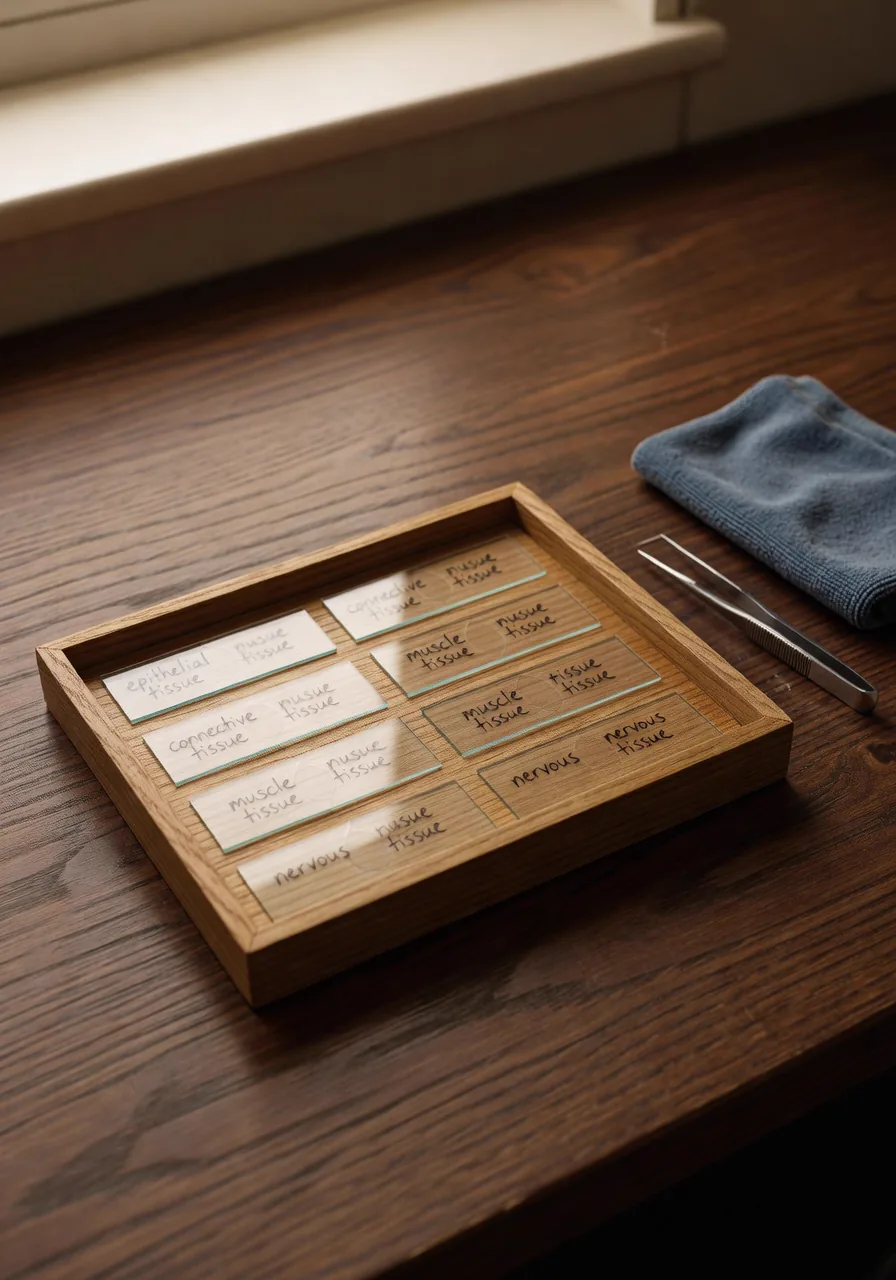
Every biology student has seen the textbook photograph of an onion-skin cell, crisp and labeled and perfectly lit. Then they sit down at a real microscope, put a real slide on the stage, and discover that the real thing is faint, off-center, out of focus, and surrounded by air bubbles and debris that look just as important as the cells. The gap between the photograph and the eyepiece is enormous, and closing it is a genuine skill.
That is why the course assesses timed microscopy identification on its own. A student is given a slide and a short list of structures to find, focus, and name — with the clock running. It sounds small. It is one of the most honest tests of competence in the whole course.
Why the clock
The time limit isn't there to add stress for its own sake. It's there because speed reveals automaticity. A student who has truly internalized how to drive a microscope (coarse focus on low power, center the target, increase magnification, fine focus, adjust the light) does it without thinking. A student who memorized the steps but never built the habit freezes. The clock separates the two cleanly, the way it does for any real skill.
You can't search the answer. The slide is right there, and either you can find the cell or you can't.
What it's really teaching
- Instrument fluency. The microscope becomes an extension of the eye, not a puzzle to solve each time.
- Visual discrimination. Telling signal from artifact — a real nucleus from a bubble, a cell wall from a scratch.
- Composure. Working accurately while a clock runs is a transferable skill that shows up everywhere from the SAT to a clinical rotation.
Like the dissection defense, this demonstration is impossible to outsource. No tool can find the cell on the student's slide for them. The microscopy rubric lays out exactly what counts as a successful identification, and the pre-lab checklist is how a student gets ready for it. The skill is small, specific, and real. Those are the best kind to assess.