Unit 01 · Plant Cells & Tissues
This unit builds from the plant cell outward: the organelles every cell shares, the three features that set plant cells apart — the cellulose cell wall, the large central vacuole, and the plastids — and how specialized cells group into the dermal, ground, and vascular tissue systems that meristems generate. Mastery means you can read a section of plant tissue under the scope as living structure with a job, not a labeled diagram to memorize.
Student learning: Connect visible plant structures to function and honest measurements
Choose the level by readiness, not age alone, and record it before instruction. Foundation, core, and honors tasks are study pathways, not an AP course or a promise of college credit. The instructor retains practical assessment and the published science rubric; integration is reported separately.
Prerequisites: Cell structure, length and area units, ratios, and the difference between an observation and a labeled model.
Suggested sequence: read and discuss the explanation; attempt the worked model; analyze the data at your selected level; check the answers; then complete the source-linked response and a fresh transfer question. These activities supplement, not replace, supervised practical work and the full-year schedule.
Assigned reading and focus
- OpenStax Biology 2e, 30.1: The Plant Body. Read Plant Organ Systems and Plant Tissues; identify dermal, ground, vascular, and meristematic tissues in the figures. Separate what a diagram labels from what an ordinary microscope resolves.
- OpenStax Biology 2e, 30.4: Leaves. Read Leaf Structure and Function and examine the stomatal diagram. Distinguish a pore, guard cells, and the surrounding epidermis.
Learn the science
Plant cells have a selectively permeable membrane, a cellulose-rich wall, plastids, and usually a large central vacuole. Water uptake can generate turgor against the wall. A cell wall and a cell membrane are different structures.
Not every plant cell is green. Plastid type depends on tissue and development: chloroplasts support photosynthesis, amyloplasts store starch, and other plastids have different roles. Many underground root cells lack chloroplasts, but exposed roots and other exceptions make an absolute rule inappropriate.
Light microscopy can reveal cell outlines, some nuclei, chloroplasts, or stomata in an appropriate preparation. It cannot identify every major organelle reliably. An epidermal peel is not a complete section through the leaf and need not show xylem.
Counts become densities only when the observed area is known. Multiple fields from one leaf improve that leaf estimate; they are not independent plants or a representative sample of an entire species.

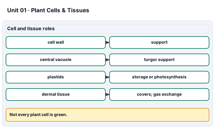
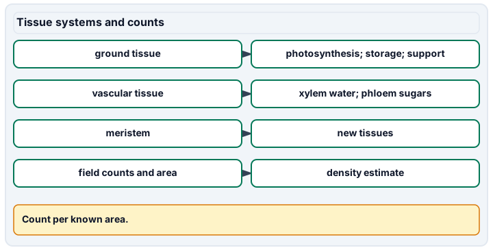
Diagram description
Plant cells have walls, plastids, and often a large central vacuole. Dermal tissue covers and controls exchange, ground tissue supports photosynthesis, storage, and support, vascular tissue includes xylem for water and phloem for sugars, and meristems generate new tissues. Counts become densities only with field area.
Data, provenance, and assumptions
| Leaf surface | Field area (mm^2) | Stomata counted |
|---|---|---|
| Lower | 0.25 | 20 |
| Lower | 0.25 | 22 |
| Lower | 0.25 | 18 |
| Lower | 0.25 | 20 |
| Upper | 0.25 | 3 |
| Upper | 0.25 | 5 |
| Upper | 0.25 | 4 |
| Upper | 0.25 | 4 |
Worked model
The lower fields contain 80 stomata in a combined 1.00 mm^2, giving 80 stomata/mm^2. The upper fields contain 16 in the same total area. The lower/upper density ratio is 5 for this leaf, not necessarily for every plant.
Numerical calibration
- 80 stomata/mm^2
- 16 stomata/mm^2
- 5 lower/upper density
Attempt the assigned level
Try the tasks before reading the calibration. These are practice answers, not a secure examination; use a new dataset or changed assumption for the assessed transfer.
Foundation
- Use the assigned diagram to explain the jobs of a cell wall, vacuole, guard cell, and stoma.
- Total the counts for each surface and identify which has the greater density.
Check after your attempt
- The wall supports and constrains expansion; the vacuole contributes to storage and turgor; guard cells regulate the stomatal pore.
- The totals are 80 and 16 over equal combined areas; the lower surface has the greater density in this record.
High-school core: typically grades 9-10
- Calculate both densities and their ratio with units.
- State the number of independent plants represented and one additional sample needed for a species-level claim.
Check after your attempt
- Lower: 80/mm^2; upper: 16/mm^2; ratio: 5.
- There is one plant. Sample multiple independently chosen plants and appropriate leaves, retaining the within-plant grouping.
Honors extension: typically grades 11-12
- Calculate the density range among fields for each surface.
- For cubic cell models with side lengths 10 and 20 micrometres, compare surface-area-to-volume ratios and state a limitation of the shape model.
Check after your attempt
- Lower fields range from 72 to 88/mm^2; upper fields from 12 to 20/mm^2.
- The ratios are 6/10 = 0.6 and 6/20 = 0.3 per micrometre. Real cell shape, membrane folds, and tissue context are not represented by perfect cubes.
History, reading, and writing connection
Use the assigned tissue figures and the historical role of microscopy to explain how an instrument changes the evidence available. Identify a feature directly visible in an approved image and one that would remain a model-based inference.
Write in your own words or use an approved accessible equivalent. Cite a specific assigned section or figure, identify its evidence, and state one limitation or counterargument. Use the AI practice contract only for permitted coaching, never to invent observations or write the assessed response.
Transfer to a new case
A new field contains 20 stomata but covers 0.50 mm^2. Is its density the same as the first lower field?
Calibration: No. Its density is 40/mm^2, half the first field's 80/mm^2. A count without an area is insufficient.
Evidence to retain
Retain the annotated figure, density calculations, sampling-unit explanation, and a limitation. Supplied counts do not certify slide preparation or microscope handling.
Record units, calculations, source/date, uncertainty, and what is measured versus inferred. A simulation or supplied dataset must stay labeled as such. Use the approved normal-care observation plan or supplied data. Do not culture unknown microbes, inoculate plants, apply hormones or pesticides, or eat study specimens.
Return to all eight learning pathways. Print this unit page for the student lessons; the linked five-page packet remains the separate assessment companion.
Unit mastery rubric
| Criterion | Developing | Proficient | Mastery |
|---|---|---|---|
| Plant cell structure & organelles | Confuses structures or claims details the image cannot resolve. | Names structures but blurs observations and diagram labels. | Explains plant-cell structures and functions, identifying only what the preparation resolves and using labeled references for other organelles. |
| Cell wall & central vacuole | Thinks the cell wall and cell membrane are the same thing. | Defines the cell wall but cannot explain turgor or the vacuole's role. | Explains how the cellulose wall and a turgid central vacuole give a plant cell its shape and support, and predicts what happens when the cell loses water. |
| Plastids & chloroplasts | Thinks every plant cell is green and full of chloroplasts. | Names plastids but treats their distribution as universal. | Distinguishes chloroplasts, chromoplasts, and amyloplasts and relates their distribution to tissue, development, and exceptions. |
| Tissue systems & meristems | Cannot name the plant tissue systems. | Lists dermal, ground, and vascular tissue but cannot place them in a real section. | Locates dermal, ground, and vascular tissue in a stem or leaf section and explains how meristems generate each. |
| Lab technique (microscopy of plant tissue) | Cannot prepare or focus the approved sample. | Observes features but misidentifies tissue or sampling area. | Uses approved peels or sections, identifies appropriate visible tissues, and calculates field density without treating fields as independent plants. |
| Integration (cross-domain) | Makes no supported connection between the source and the science. | Uses the source but needs help connecting evidence, writing, or limitations to the science. | Independently connects History, Reading, and Writing using a cited source, appropriate evidence, a limitation, and a scientific explanation. |
Integration is reported separately and cannot lower the science grade or block a science demonstration pass. Science and practical criteria determine that pass. Use the integration guide's evidence checklist for the separately reported criterion.
“A plant cell has a cellulose wall and a big central vacuole an animal cell doesn’t — when the vacuole is full of water the cell is turgid and holds the leaf firm; when it dries out the plant wilts. The green is chloroplasts, but the root cells I looked at had none, because they aren’t doing photosynthesis.”
“It’s got a wall… and it’s green because plants are green? I found the cells but I’m not sure which part is which.”
You demonstrate this unit through microscopy labs — epidermal peels and prepared cross-sections — plus short oral checks where you reason from what you see aloud, not a multiple-choice test. A criterion counts as mastered only when you can both run the technique and justify the plant biology behind it. Mastery is demonstrated, not awarded.
A 5-page clipboard packet — unit overview, key terms, the mastery rubric, anchor examples, and a score sheet you can print and grade against.