Unit 01 · The Microscope: Parts, Care & Focusing
This is the first rung of the ladder, and every rung above it depends on this one. You learn the compound microscope by using it: naming each part and what it does, carrying and storing it without damage, setting illumination and the diaphragm for contrast, and focusing cleanly — low power first, coarse then fine, always moving away from the slide. Mastery here is not something you can explain your way into. An instructor watches you do it, and the image in the eyepiece is the proof.
Student learning: Operate the microscope as an optical system, not a mystery box
Choose the level by readiness, not age alone, and record it before instruction. Foundation, core, and honors tasks are study pathways, not an AP course or a promise of college credit. The instructor retains practical assessment and the published science rubric; integration is reported separately.
Prerequisites: Basic ratio language, careful handling, and the published Microscope parts/care rubric.
Suggested sequence: read and discuss the explanation; attempt the worked model; analyze the data at your selected level; check the answers; then complete the source-linked response and a fresh transfer question. These activities supplement, not replace, supervised practical work and the semester schedule.
Assigned reading and focus
- OpenStax Microbiology, 2.3: Instruments of Microscopy. Read Brightfield Microscopes and Microscope Maintenance: identify ocular, objective, condenser, diaphragm, stage, coarse focus and fine focus. Record how objective magnification and ocular magnification combine.
Learn the science
A compound microscope uses the ocular lens and one selected objective together. The objective may be 4x, 10x, 40x or 100x; the total magnification is objective power multiplied by the ocular power, commonly 10x.
Care is part of the science evidence. A dirty, scratched or crashed lens changes the image before interpretation begins. Low power first, coarse then fine focus, and lens paper only are not arbitrary habits.
Supplied-image paper work can assess reasoning, scale and evidence limits; hands-on technique is assessed only when directly observed or through an approved accessible equivalent. A student can calculate totals on paper, but carrying, focusing and care are mastered only when the educator observes the technique.

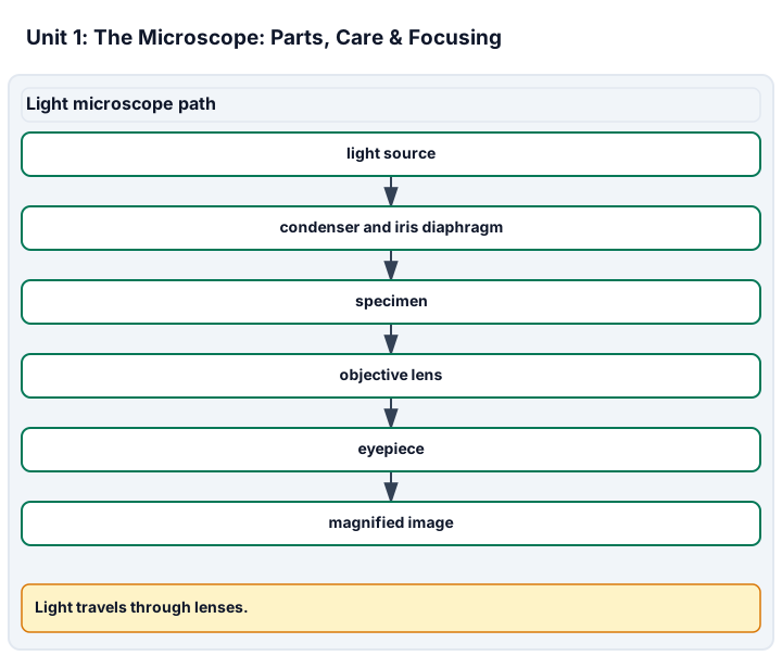
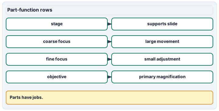
Diagram description
The light path runs from light source to condenser and iris diaphragm, specimen, objective lens, eyepiece and magnified image. The grid states stage supports the slide, coarse focus makes large movements, fine focus makes small adjustments, and the objective provides primary magnification.
Data, provenance, and assumptions
| Objective label | Objective power | Ocular power | Total magnification |
|---|---|---|---|
| Scanning | 4 | 10 | 40 |
| Low | 10 | 10 | 100 |
| High dry | 40 | 10 | 400 |
| Oil immersion | 100 | 10 | 1000 |
Worked model
A 40x objective with a 10x ocular gives 400x total magnification. A 100x objective with the same ocular gives 1000x total; calling that total an objective power confuses the part with the whole optical system.
Numerical calibration
- 400 total magnification
- 1000 total magnification
Attempt the assigned level
Try the tasks before reading the calibration. These are practice answers, not a secure examination; use a new dataset or changed assumption for the assessed transfer.
Foundation: typically grades 7-8
- Match each lens and focus control to its job using the assigned source.
- Calculate the total magnification for the 4x and 10x objectives.
Check after your attempt
- The ocular is the eyepiece, the objective is the lens near the slide, the condenser focuses light, and the diaphragm controls light reaching the specimen.
- 4x times 10x is 40x total; 10x times 10x is 100x total.
High-school core: typically grades 9-10
- Calculate every total in the table and explain why care affects evidence.
- Write the focusing sequence and name which step must be observed by the educator.
Check after your attempt
- The totals are 40x, 100x, 400x and 1000x. Lens care matters because dirt, scratches or a crashed lens degrade the specimen image.
- Start low power, use coarse then fine focus, center the target, and move to higher objectives using fine focus only. The hands-on focusing technique must be observed.
Honors/readiness extension: quantitative calibration and image-integrity reasoning
- Explain why a higher objective is not automatically a better scientific image.
- Compare a calculation-only response with an observed practical demonstration.
Check after your attempt
- Higher magnification can dim the image, narrow the field and expose preparation or resolution limits; clarity requires alignment, light and resolving power.
- The calculation shows optical understanding. It does not certify carrying, stage control, focusing or slide safety without observation.
History, reading, and writing connection
Use OpenStax Biology 4.1 only for its stated history: Hooke used Micrographia to name cork chambers cells, and van Leeuwenhoek observed single-celled organisms. Connect that source-supported history to the instrument limits a student must record today; do not invent quotations or motives.
Write in your own words or use an approved accessible equivalent. Cite a specific assigned section or figure, identify its evidence, and state one limitation or counterargument. Use the AI practice contract only for permitted coaching, never to invent observations or write the assessed response.
Transfer to a new case
A student writes the total magnification as if it were the objective label after using a 100x oil objective and a 10x ocular. What correction belongs in the record?
Calibration: Write "100x objective with 10x ocular = 1000x total magnification." The objective label and total magnification are different quantities.
Evidence to retain
Retain the lens-total calculation, focusing sequence, approved practical observation record or alternative, and the corrected objective-vs-total language.
Record units, calculations, source/date, uncertainty, and what is measured versus inferred. A simulation or supplied dataset must stay labeled as such. Use only educator-approved slides, specimens, stains, reagents and images. Review SDS information before approved stains, handle slides and coverslips through the approved sharps procedure, and do not culture, amplify or improvise work with unknown microorganisms.
Return to all eight learning pathways. Print this unit page for the student lessons; the linked five-page packet remains the separate assessment companion.
Evidence boundary for this unit
Paper calculations can show objective-vs-total understanding. The actual microscope-care and focusing criteria require observed technique with approved equipment or a stated accessible equivalent.
Retain the lens-total calculation, focusing sequence, approved practical observation record or alternative, and the corrected objective-vs-total language.
- Science 1: Parts & their jobs. Lens labels, total-magnification calculation and part-function explanation.
- Science 4: Focusing procedure. Observed low-power-first focusing or an approved accessible equivalent observed by the educator.
Course investigation inventory. Completion evidence is a calibrated, honestly documented image or drawing, plus an independently repeatable size or count estimate when the unit calls for one. Supplied-image paper work is distinct from an observed microscope practical.
Unit mastery rubric
| Criterion | Developing | Proficient | Mastery |
|---|---|---|---|
| Parts & their jobs | Cannot reliably name the objectives, condenser, diaphragm, or focus knobs, or say what each does. | Names most parts but confuses coarse vs. fine focus, or what the diaphragm changes. | Names each microscope part, distinguishes objective power from total magnification, and explains how each part shapes the image. |
| Carrying, storage & care | Grabs the scope one-handed or leaves it on high power; wipes lenses with a shirt or paper towel. | Carries and stores it correctly when reminded, but forgets to return to low power or to use lens paper. | Carries with two hands (arm and base), stores on the lowest objective with the cord wrapped, and cleans optics only with lens paper. |
| Illumination & contrast | Leaves the light at one setting and ignores the diaphragm; the image is washed out or too dark to read. | Adjusts brightness but not the diaphragm, so faint specimens stay invisible. | Sets the lamp and works the iris diaphragm to bring out contrast without glare, retuning it for each specimen. |
| Focusing procedure | Focuses down toward the slide on high power — risking a cracked slide or a scratched objective. | Starts on low power but reaches for the coarse knob on high power, or racks the wrong direction. | Performs low-power-first focusing and fine-focus-only transitions (observed) while explaining how the sequence protects the slide and lens. |
| Centering, scanning & the mechanical stage | Raises magnification before finding or centering the specimen and loses it off the field. | Centers on low power but drives the stage jerkily and drifts off the target. | Finds and centers the specimen on low power first, then scans smoothly with the mechanical stage as magnification climbs. |
| Integration (cross-domain) | Makes no supported connection between the source and the science. | Uses the source but needs help connecting evidence, writing, or limitations to the science. | Independently connects History, Reading, and Writing using a cited source, appropriate evidence, a limitation, and a scientific explanation. |
Integration is reported separately and cannot lower the science grade or block a science demonstration pass. Science and practical criteria determine that pass. Use the integration guide's evidence checklist for the separately reported criterion.
I used the 40x objective with a 10x ocular, so I recorded 400x total. I focused from low power and switched to fine focus only before moving up, because the record needs both the optical calculation and the safe technique.
I used the biggest lens and called it 1000x. I turned whichever knob made the image appear and did not record the ocular or objective.
Paper calculations can show objective-vs-total understanding. The actual microscope-care and focusing criteria require observed technique with approved equipment or a stated accessible equivalent.
A 5-page clipboard packet — unit overview, key terms, the mastery rubric, anchor examples, and a score sheet you can print and grade against.