Unit 02 · Magnification, Resolution & Measurement
Once you can focus a scope, the next rung is knowing what you are actually looking at — how big it is and how much detail you can trust. You learn to compute total magnification from the ocular and objective, to tell resolution and field of view apart from raw power, to measure the diameter of the field, and to estimate a specimen's real size from what fills it. An instructor watches you calibrate an ocular scale against a stage micrometer and read a measurement off the eyepiece — the number you report is the proof.
Student learning: Measure with calibration and state what resolution can actually separate
Choose the level by readiness, not age alone, and record it before instruction. Foundation, core, and honors tasks are study pathways, not an AP course or a promise of college credit. The instructor retains practical assessment and the published science rubric; integration is reported separately.
Prerequisites: Ratios, decimals, micrometres, averages, range, and Unit 01 objective-vs-total language.
Suggested sequence: read and discuss the explanation; attempt the worked model; analyze the data at your selected level; check the answers; then complete the source-linked response and a fresh transfer question. These activities supplement, not replace, supervised practical work and the semester schedule.
Assigned reading and focus
- OpenStax Microbiology, 2.3: Instruments of Microscopy. Read the total magnification formula and the note that magnifying further can enlarge without increasing resolution.
- Nikon MicroscopyU: Numerical Aperture. Read how numerical aperture describes the light cone an objective can collect and why NA affects resolving power. This is an honors comparison, not a required equipment purchase.
- Nikon MicroscopyU: Resolution. Read the definition of resolution and the role of numerical aperture; treat numerical aperture as an honors extension, not a required purchase.
Learn the science
Magnification makes the image larger. Resolution separates close details. A larger blur is not more evidence.
A stage micrometer calibrates distance; an ocular scale is useful only after calibration for the selected objective. Repeated measurements expose reading spread and uncertainty.
Numerical aperture and condenser/objective matching explain why some systems resolve more detail. That comparison belongs in honors reasoning; the course does not require families to buy advanced objectives.

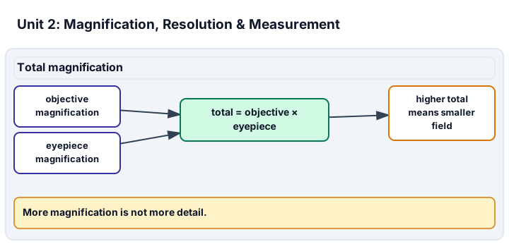
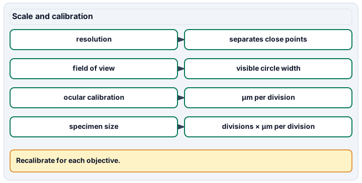
Diagram description
Total magnification uses objective and eyepiece magnification to compute total magnification and note the smaller field at higher total. Scale and calibration defines resolution, field of view, ocular calibration and specimen size formulas.
Data, provenance, and assumptions
| Total magnification | Field diameter (mm) | Specimen fraction of field |
|---|---|---|
| 40 | 4.5 | 0.036 |
| 100 | 1.8 | 0.09 |
| 400 | 0.45 | 0.36 |
| Trial | Micrometres per 10 ocular divisions |
|---|---|
| A | 102 |
| B | 99 |
| C | 101 |
Worked model
At 400x total, the field is 0.45 mm = 450 micrometres. A specimen spanning 0.36 of the field is 162 micrometres. The repeated ocular-scale readings average about 100.7 micrometres per 10 divisions, with a 3 micrometre full range.
Numerical calibration
- 162 micrometres
- 100.66666666666667 micrometres per 10 divisions
- 1.5 micrometres
Attempt the assigned level
Try the tasks before reading the calibration. These are practice answers, not a secure examination; use a new dataset or changed assumption for the assessed transfer.
Foundation: typically grades 7-8
- Define magnification and resolution in separate sentences.
- Use the table to estimate the specimen size at 400x total.
Check after your attempt
- Magnification enlarges appearance; resolution separates close details.
- 0.45 mm is 450 micrometres; 0.36 of that is 162 micrometres.
High-school core: typically grades 9-10
- Calculate the repeated-calibration mean and report a simple half-range uncertainty.
- Explain why nominal magnification alone is not enough for a size claim.
Check after your attempt
- The mean is about 100.7 micrometres per 10 divisions; half the range is 1.5 micrometres.
- The same nominal magnification can have different field diameters or resolution, so calibration and setup must be named.
Honors/readiness extension: quantitative calibration and image-integrity reasoning
- Use the Nikon source to explain why numerical aperture affects resolving power.
- Compare the uncertainty from the ocular repeats with the field-fraction estimate.
Check after your attempt
- Higher numerical aperture collects a wider cone of light and improves resolving power under matched conditions.
- The ocular repeat spread belongs to the calibration; the field-fraction estimate also depends on judging the specimen edge. Both limits should be reported.
History, reading, and writing connection
History: OpenStax Microbiology 2.3 opens by noting that Joseph Jackson Lister created an essentially modern light microscope in 1830 and that later advances improved magnification, resolution and contrast. Use the OpenStax and Nikon readings to explain how an instrument claim needs both scale and resolution, and why even a better instrument still needs calibration. State that numerical aperture is an extension for comparing imaging methods, not a purchase requirement.
Write in your own words or use an approved accessible equivalent. Cite a specific assigned section or figure, identify its evidence, and state one limitation or counterargument. Use the AI practice contract only for permitted coaching, never to invent observations or write the assessed response.
Transfer to a new case
A supplied image has a 200 micrometre field diameter and a specimen spans one quarter of it. What size estimate and limit belong in the record?
Calibration: The estimate is 50 micrometres. The record should name the calibration source and say edge judgment and calibration uncertainty limit the estimate.
Evidence to retain
Retain the calibrated field diameter, size calculation, repeated-measurement spread, and a resolution-vs-magnification statement.
Record units, calculations, source/date, uncertainty, and what is measured versus inferred. A simulation or supplied dataset must stay labeled as such. Use only educator-approved slides, specimens, stains, reagents and images. Review SDS information before approved stains, handle slides and coverslips through the approved sharps procedure, and do not culture, amplify or improvise work with unknown microorganisms.
Return to all eight learning pathways. Print this unit page for the student lessons; the linked five-page packet remains the separate assessment companion.
Evidence boundary for this unit
The size estimate can be assessed from supplied images. Stage-micrometer handling and microscope alignment are practical evidence only when observed.
Retain the calibrated field diameter, size calculation, repeated-measurement spread, and a resolution-vs-magnification statement.
- Science 2: Magnification vs. resolution. Written distinction and an example where magnification does not add detail.
- Science 5: Calibrating with a stage micrometer. Observed stage-micrometer calibration, plus the repeated record with mean, units and uncertainty.
Course investigation inventory. Completion evidence is a calibrated, honestly documented image or drawing, plus an independently repeatable size or count estimate when the unit calls for one. Supplied-image paper work is distinct from an observed microscope practical.
Unit mastery rubric
| Criterion | Developing | Proficient | Mastery |
|---|---|---|---|
| Total magnification (ocular × objective) | Reads only the objective number and calls it the magnification, or cannot say where the two numbers come from. | Multiplies ocular by objective when reminded but forgets the ocular power or reads the wrong objective. | Reads the ocular and objective powers off the scope and states the total magnification for each objective on sight. |
| Magnification vs. resolution | Assumes the highest-power objective always gives the best view. | Knows resolution and field of view matter but still reaches for maximum power first. | Distinguishes enlargement from resolving power and states when extra magnification no longer adds usable detail. |
| Measuring field-of-view diameter | Cannot say how wide the field is at any objective. | Measures the field on low power but cannot work out the others. | Measures the field diameter against a ruler on low power and calculates it for each higher objective from the magnification change. |
| Estimating specimen size from the field | Guesses a specimen's size with nothing to compare it to. | Estimates size but ignores how much of the field the specimen fills. | Estimates a specimen's real size from the fraction of the known field it spans and reports it in sensible units. |
| Calibrating with a stage micrometer | Uses the eyepiece scale as if its marks were fixed distances. | Lines up the stage micrometer but cannot convert eyepiece units into real distance. | Calibrates the ocular or field measurement with a stage micrometer (observed), repeats readings, and reports an uncertainty rather than a single naked number. |
| Integration (cross-domain) | Makes no supported connection between the source and the science. | Uses the source but needs help connecting evidence, writing, or limitations to the science. | Independently connects History, Reading, and Writing using a cited source, appropriate evidence, a limitation, and a scientific explanation. |
Integration is reported separately and cannot lower the science grade or block a science demonstration pass. Science and practical criteria determine that pass. Use the integration guide's evidence checklist for the separately reported criterion.
Assigned practice: calibrate the ocular scale, not the reference ruler
Read the brightfield magnification discussion in OpenStax Microbiology, Instruments of Microscopy. Magnification and resolution differ. An ocular reticle has arbitrary divisions until compared with a known stage micrometer at the current objective and optical configuration.
Model: micrometres per ocular division = known stage length divided by aligned ocular divisions. Specimen size = that calibration factor times the observed specimen divisions. Recalibrate for each objective or changed optical configuration; enlarging an image does not create additional resolved detail.
| Objective | Known stage span (micrometres) | Matching ocular divisions | Specimen divisions |
|---|---|---|---|
| 10 | 100 | 10 | 20 |
| 40 | 100 | 40 | 80 |
Foundation: explain why the ocular divisions do not have one fixed physical size. Core: calculate both calibration factors and both specimen sizes. Honors: explain the error caused by using the 10x factor on the 40x reading, and identify reference, alignment, boundary and resolution uncertainty.
Evidence: retain calculations, objective/configuration, units, and an uncertainty explanation. Transfer: 50 micrometres matching 25 divisions gives 2 micrometres/division. Supplied calculations demonstrate measurement reasoning; an instructor separately observes actual calibration technique.
My 400x-total field is calibrated at 450 micrometres, so the specimen at 0.36 field is about 162 micrometres. The ocular repeat mean is 100.7 micrometres per 10 divisions, with 1.5 micrometres half-range uncertainty.
It was on high power, so I said the specimen was definitely tiny and did not measure the field or repeat the scale.
The size estimate can be assessed from supplied images. Stage-micrometer handling and microscope alignment are practical evidence only when observed.
A 5-page clipboard packet — unit overview, key terms, the mastery rubric, anchor examples, and a score sheet you can print and grade against.