Unit 03 · Preparing Wet Mounts
A good image starts before you ever look through the eyepiece — it starts with the mount. On this rung you learn to handle a clean slide without smudging it, lay down a specimen thin enough to see through, add just the right drop of water, and lower the coverslip at a shallow angle so no air bubbles get trapped. An instructor watches you build the mount and then read your own field: mastery is a clear, bubble-free view, not a description of one.
Student learning: Separate specimen variation from preparation artifacts
Choose the level by readiness, not age alone, and record it before instruction. Foundation, core, and honors tasks are study pathways, not an AP course or a promise of college credit. The instructor retains practical assessment and the published science rubric; integration is reported separately.
Prerequisites: Slide/coverslip safety, Unit 01 focusing, thin specimens, and the approved wet-mount procedure.
Suggested sequence: read and discuss the explanation; attempt the worked model; analyze the data at your selected level; check the answers; then complete the source-linked response and a fresh transfer question. These activities supplement, not replace, supervised practical work and the semester schedule.
Assigned reading and focus
- OpenStax Biology 2e, 4.1: Studying Cells. Read Light Microscopes: living organisms can be viewed with light microscopes, while transparent cells often need contrast; connect that claim to wet-mount thickness and artifacts.
Learn the science
A wet mount is a preparation, not the specimen itself. Bubbles, folds, flooded edges and thick tissue can create artifacts that look more dramatic than the actual cells.
Specimen variation is real: cells can differ by tissue, age, orientation and preparation. The record must separate what was observed from what the preparation may have introduced.
No improvised procedures: use approved specimens, approved images, SDS-reviewed stains and the assigned sharps procedure. Unknown microbes are never cultured, amplified or collected for this course. The learning pathway may use approved images with the same conceptual standard; it cannot certify slide handling without observation.

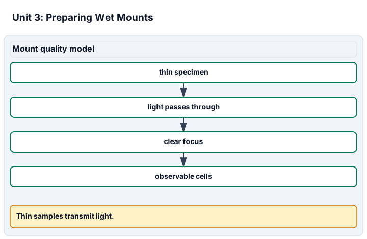
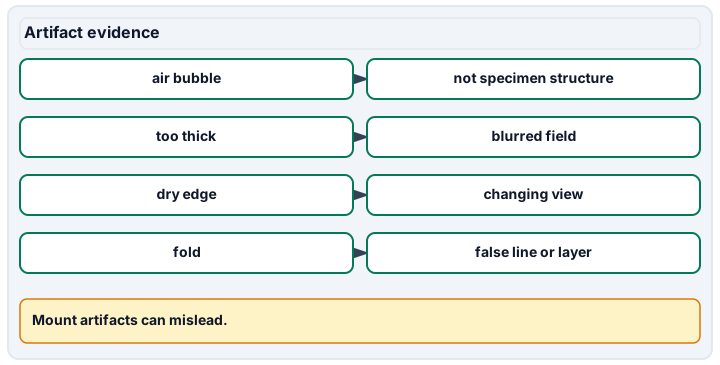
Diagram description
Mount quality model links a thin specimen to transmitted light, clear focus and observable cells. Artifact evidence distinguishes air bubbles, thick samples, dry edges and folds from actual specimen structures.
Data, provenance, and assumptions
| Attempt | Bubbles in field | Visible cells | Remake needed |
|---|---|---|---|
| A | 0 | 42 | no |
| B | 5 | 12 | yes |
| C | 1 | 38 | no |
| D | 4 | 10 | yes |
| E | 0 | 40 | no |
Worked model
Two of five attempts need remaking, a 40% remake rate. The record does not treat bubbles as organisms or hidden cells; it says which fields were artifacts of preparation.
Numerical calibration
- 40 percent of attempts remade
- 40 visible cells in non-remake fields
Attempt the assigned level
Try the tasks before reading the calibration. These are practice answers, not a secure examination; use a new dataset or changed assumption for the assessed transfer.
Foundation: typically grades 7-8
- List two artifacts that can come from preparation rather than the specimen.
- Calculate the remake fraction.
Check after your attempt
- Bubbles and folds are preparation artifacts; excess water or a thick specimen can also obscure cells.
- 2 of 5 attempts needed remaking, so the fraction is 0.40 or 40%.
High-school core: typically grades 9-10
- Explain how you would distinguish specimen variation from a bubble artifact using the supplied record.
- State why approved image work is different from a practical slide-prep assessment.
Check after your attempt
- A bubble has an edge/rim and may shift differently from tissue; repeat or remake records help show whether the feature persists.
- Images can assess interpretation and artifact recognition; handling slides and coverslips must be watched.
Honors/readiness extension: quantitative calibration and image-integrity reasoning
- Design a repeated-measurement record for comparing two approved specimen or image sources without changing the procedure.
- Name one access alternative that keeps the same conceptual standard.
Check after your attempt
- Use the same approved mounting procedure, record source, attempt, field, artifact count and cell visibility, and compare fields without treating fields as independent specimens.
- Use supplied images or virtual microscopy with artifact flags and require honest evidence limits; do not claim hands-on technique unless observed.
History, reading, and writing connection
History: OpenStax Biology 4.1 reports that van Leeuwenhoek, skilled in crafting lenses, observed the movements of single-celled organisms he called "animalcules." Use the OpenStax light-microscope passage to explain why a transparent or living specimen can be hard to interpret. Connect the source to one preparation artifact and one honest record limit.
Write in your own words or use an approved accessible equivalent. Cite a specific assigned section or figure, identify its evidence, and state one limitation or counterargument. Use the AI practice contract only for permitted coaching, never to invent observations or write the assessed response.
Transfer to a new case
A new image shows a perfect dark ring crossing several cells. What should the learner record before identifying it as a structure?
Calibration: Record it as a possible bubble or preparation artifact, compare another field or remake record, and avoid naming it as a cell structure until evidence supports that claim.
Evidence to retain
Retain the approved source, artifact notes, remake decision, image or observed field, and whether hands-on prep was actually observed.
Record units, calculations, source/date, uncertainty, and what is measured versus inferred. A simulation or supplied dataset must stay labeled as such. Use only educator-approved slides, specimens, stains, reagents and images. Review SDS information before approved stains, handle slides and coverslips through the approved sharps procedure, and do not culture, amplify or improvise work with unknown microorganisms.
Return to all eight learning pathways. Print this unit page for the student lessons; the linked five-page packet remains the separate assessment companion.
Evidence boundary for this unit
Conceptual artifact recognition can use supplied images. Clean slide handling, coverslip lowering and wicking remain observed practical criteria.
Retain the approved source, artifact notes, remake decision, image or observed field, and whether hands-on prep was actually observed.
- Science 2: Placing a thin specimen. Observed specimen placement, plus thickness/artifact notes.
- Science 5: Wicking & fixing a bad mount. Observed wicking/remake decision, plus what changed between attempts.
Course investigation inventory. Completion evidence is a calibrated, honestly documented image or drawing, plus an independently repeatable size or count estimate when the unit calls for one. Supplied-image paper work is distinct from an observed microscope practical.
Unit mastery rubric
| Criterion | Developing | Proficient | Mastery |
|---|---|---|---|
| Clean slide handling | Grabs the slide by its faces, leaving fingerprints and smears across the viewing area. | Holds the slide by the edges when reminded but forgets to check it is clean and dry first. | Handles the slide by its edges only, checks it is clean and dry, and keeps the viewing area free of prints and dust. |
| Placing a thin specimen | Piles on a chunk of specimen too thick for light to pass through. | Chooses a thin piece but centers it poorly or lets it fold over. | Selects or peels a thin specimen, lays it flat and centered (observed), and separates specimen variation from artifacts. |
| Sizing the water drop | Floods the slide or adds no water at all. | Adds water but consistently too much or too little for the coverslip. | Places a single drop sized so it spreads just under the coverslip without overflowing or leaving dry gaps. |
| Lowering the coverslip | Drops the coverslip flat, trapping a field full of air bubbles. | Lowers it at an angle sometimes but still leaves stray bubbles. | Rests one edge of the coverslip in the water and lowers it slowly at a shallow angle so the water spreads bubble-free. |
| Wicking & fixing a bad mount | Leaves excess water pooling or gives up when the mount goes wrong. | Wicks excess water but cannot diagnose why a mount looks bad. | Wicks excess and remakes a bad mount (observed), recording what changed without treating artifacts as organisms. |
| Integration (cross-domain) | Makes no supported connection between the source and the science. | Uses the source but needs help connecting evidence, writing, or limitations to the science. | Independently connects History, Reading, and Writing using a cited source, appropriate evidence, a limitation, and a scientific explanation. |
Integration is reported separately and cannot lower the science grade or block a science demonstration pass. Science and practical criteria determine that pass. Use the integration guide's evidence checklist for the separately reported criterion.
I marked B and D for remake because bubbles and low visible-cell counts make the field unreliable. That is a preparation-artifact note, not a claim that the specimen had ring-shaped cells.
I counted every circle as a cell and kept the flooded slide because the image looked interesting.
Conceptual artifact recognition can use supplied images. Clean slide handling, coverslip lowering and wicking remain observed practical criteria.
A 5-page clipboard packet — unit overview, key terms, the mastery rubric, anchor examples, and a score sheet you can print and grade against.