Unit 04 · Staining & Contrast Techniques
Many specimens are nearly transparent until you add contrast. On this rung you learn to pick the right stain for what you are viewing, draw it through the mount without flooding it, and tune the light so faint structures stand out. An instructor watches you stain a mount and compare it against an unstained one: mastery is a field where the detail finally shows, not an explanation of why it should.
Student learning: Use approved contrast choices and document how they changed the image
Choose the level by readiness, not age alone, and record it before instruction. Foundation, core, and honors tasks are study pathways, not an AP course or a promise of college credit. The instructor retains practical assessment and the published science rubric; integration is reported separately.
Prerequisites: Wet-mount artifact recognition, SDS-reviewed stain handling, and basic difference calculations.
Suggested sequence: read and discuss the explanation; attempt the worked model; analyze the data at your selected level; check the answers; then complete the source-linked response and a fresh transfer question. These activities supplement, not replace, supervised practical work and the semester schedule.
Assigned reading and focus
- OpenStax Biology 2e, 4.1: Studying Cells. Read Light Microscopes: cells are often transparent and stains may be needed to distinguish components; staining usually changes or kills cells.
Learn the science
Staining is an approved contrast decision, not a decoration. The record names the stain, source, SDS/approval status, exposure and what changed in the image.
Contrast can also come from diaphragm settings and illumination. The learner should compare stained and unstained fields without claiming the stain revealed every structure.
No improvised procedures: use approved specimens, approved images, SDS-reviewed stains and the assigned sharps procedure. Unknown microbes are never cultured, amplified or collected for this course. Stain choices and disposal stay with the educator-approved procedure.

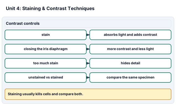
Diagram description
Contrast controls states that stain can absorb light and add contrast, closing the iris diaphragm can increase contrast while reducing light, too much stain hides detail, and unstained and stained observations should compare the same specimen.
Data, provenance, and assumptions
| Field | Background brightness | Structure brightness |
|---|---|---|
| Unstained | 20 | 18 |
| Approved stain | 20 | 11 |
| Overstained | 20 | 4 |
Worked model
The unstained background-structure difference is 2 brightness units; the approved stain difference is 9. Overstaining gives a larger difference from background but hides detail, so it is not automatically better.
Numerical calibration
- 9 brightness-unit difference
- 7 gain over unstained field
Attempt the assigned level
Try the tasks before reading the calibration. These are practice answers, not a secure examination; use a new dataset or changed assumption for the assessed transfer.
Foundation: typically grades 7-8
- Explain why stain approval and SDS review matter.
- Calculate the approved-stain contrast difference.
Check after your attempt
- Stains are chemicals and can change cells; the educator approves use, handling and disposal.
- 20 minus 11 gives a 9-unit difference.
High-school core: typically grades 9-10
- Compare unstained, approved stain and overstained records without treating maximum darkness as mastery.
- State how diaphragm adjustment differs from stain selection.
Check after your attempt
- Approved stain improved contrast; overstaining may obscure detail, so the evidence includes visibility and artifacts.
- The diaphragm changes light reaching the specimen; stain changes the specimen or its visible structures.
Honors/readiness extension: quantitative calibration and image-integrity reasoning
- Design an honest before/after image record using the same field or a matched field.
- Explain why image contrast values do not by themselves identify a structure.
Check after your attempt
- Record the source, stain, timing, illumination/diaphragm setting, field, and whether the same or matched field was used.
- Brightness differences show contrast, not biological identity; shape, location, preparation and source are still needed.
History, reading, and writing connection
History: OpenStax Biology 4.1 notes that later advances in lenses, microscope construction and staining techniques let scientists see some components inside cells. Use the OpenStax statement about stains and transparent cells to explain the trade-off: contrast may reveal components, but the preparation changes the specimen and must be documented.
Write in your own words or use an approved accessible equivalent. Cite a specific assigned section or figure, identify its evidence, and state one limitation or counterargument. Use the AI practice contract only for permitted coaching, never to invent observations or write the assessed response.
Transfer to a new case
A field is darker after extra stain, but the cell boundaries disappear. Should that be scored as better contrast?
Calibration: No. The record should say the extra stain increased darkness but reduced interpretable detail; compare with the approved stain field and redo only through the approved procedure.
Evidence to retain
Retain the approved stain/source, SDS or educator approval note, before/after comparison, diaphragm setting and artifact limit.
Record units, calculations, source/date, uncertainty, and what is measured versus inferred. A simulation or supplied dataset must stay labeled as such. Use only educator-approved slides, specimens, stains, reagents and images. Review SDS information before approved stains, handle slides and coverslips through the approved sharps procedure, and do not culture, amplify or improvise work with unknown microorganisms.
Return to all eight learning pathways. Print this unit page for the student lessons; the linked five-page packet remains the separate assessment companion.
Evidence boundary for this unit
Interpreting supplied before/after images is a paper task. Applying approved stain and managing materials safely are assessed only when observed.
Retain the approved stain/source, SDS or educator approval note, before/after comparison, diaphragm setting and artifact limit.
- Science 1: Choosing the stain. Approved stain/reagent record with reason and SDS/educator approval.
- Science 5: Comparing stained vs. unstained fields. Before/after contrast comparison with preparation limits.
Course investigation inventory. Completion evidence is a calibrated, honestly documented image or drawing, plus an independently repeatable size or count estimate when the unit calls for one. Supplied-image paper work is distinct from an observed microscope practical.
Unit mastery rubric
| Criterion | Developing | Proficient | Mastery |
|---|---|---|---|
| Choosing the stain | Reaches for whatever stain is closest with no reason for the choice. | Knows there are different stains but cannot match one to the specimen. | Uses only educator-approved stains/reagents with SDS-reviewed handling and explains why that contrast choice fits the specimen. |
| Applying stain under the coverslip | Lifts the coverslip and floods stain straight onto the specimen. | Places stain at the coverslip edge but does not draw it evenly through. | Adds a drop at one coverslip edge and wicks from the other with paper, drawing the stain evenly under without lifting the coverslip. |
| Avoiding over-staining | Soaks the specimen until the whole field is one dark color. | Uses less stain but still darkens the field past the point of useful detail. | Applies just enough stain to reveal structure, stopping before the field goes muddy, and dilutes or rinses when it does. |
| Tuning the iris diaphragm for contrast | Leaves the diaphragm wide open so a stained specimen washes out. | Adjusts brightness but not the diaphragm to bring out contrast. | Works the iris diaphragm to balance light against the stain so structures stand out without glare. |
| Comparing stained vs. unstained fields | Views only the stained slide and cannot say what the stain changed. | Looks at both but describes them only in vague terms. | Compares stained and unstained fields with the same evidence standard, naming both revealed structures and artifacts introduced by preparation. |
| Integration (cross-domain) | Makes no supported connection between the source and the science. | Uses the source but needs help connecting evidence, writing, or limitations to the science. | Independently connects History, Reading, and Writing using a cited source, appropriate evidence, a limitation, and a scientific explanation. |
Integration is reported separately and cannot lower the science grade or block a science demonstration pass. Science and practical criteria determine that pass. Use the integration guide's evidence checklist for the separately reported criterion.
The approved stain changed the structure-background difference from 2 to 9 units, but I did not score the overstained field as best because the details were obscured.
I added more stain until the field was dark and assumed the darkest image must be the most scientific.
Interpreting supplied before/after images is a paper task. Applying approved stain and managing materials safely are assessed only when observed.
A 5-page clipboard packet — unit overview, key terms, the mastery rubric, anchor examples, and a score sheet you can print and grade against.