Unit 06 · Animal Cells & Histology
Now you turn the scope on animals. You prepare your own cheek-cell wet mount — a soft cell with no wall — and then tour a set of prepared histology slides, learning to orient a thin section and name the tissue in front of you. Mastery here is not something you can explain your way into. An instructor watches you mount, orient, and identify, and the tissue under the objective is the proof.
Student learning: Read animal-cell and tissue evidence without adding medical claims
Choose the level by readiness, not age alone, and record it before instruction. Foundation, core, and honors tasks are study pathways, not an AP course or a promise of college credit. The instructor retains practical assessment and the published science rubric; integration is reported separately.
Prerequisites: Animal/plant cell comparison, approved image use, area units, and non-diagnostic language.
Suggested sequence: read and discuss the explanation; attempt the worked model; analyze the data at your selected level; check the answers; then complete the source-linked response and a fresh transfer question. These activities supplement, not replace, supervised practical work and the semester schedule.
Assigned reading and focus
- OpenStax Biology 2e, 4.1: Studying Cells. Read the opening description of cells, tissues, organs and organ systems; use it to explain why a tissue image is not a medical diagnosis.
Learn the science
Animal-cell and histology slides show prepared tissue evidence. A student can compare cell boundaries, nuclei and tissue organization without making diagnostic claims.
A supplied image or virtual microscopy set can assess pattern recognition and measurement. Preparing cheek-cell wet mounts remains an observed practical if it is assigned.
No personal health data are collected or uploaded. Use de-identified approved images, educator-provided specimens, or, only when assigned, the student's own cheek cells under the approved, observed procedure.

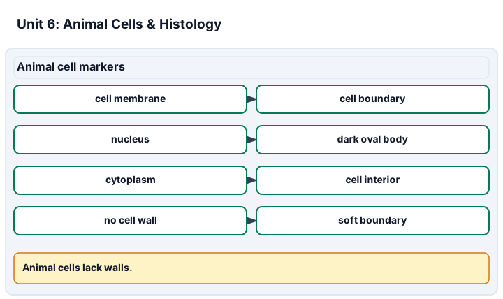
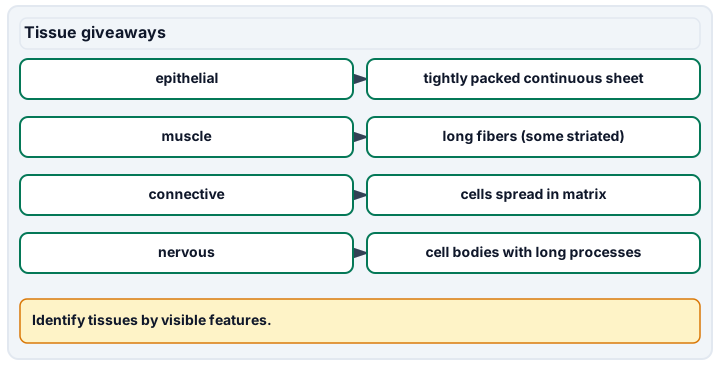
Diagram description
Animal cell markers connects membrane, nucleus, cytoplasm and lack of cell wall with microscope evidence. Tissue giveaways pairs epithelial, muscle, connective and nervous tissues with visible features.
Data, provenance, and assumptions
| Field | Area (mm^2) | Nuclei counted | Tissue pattern |
|---|---|---|---|
| A | 0.15 | 28 | epithelial layer |
| B | 0.15 | 12 | muscle fibers |
| C | 0.15 | 18 | connective tissue |
Worked model
Field A has 28 nuclei in 0.15 mm^2, about 186.7 nuclei/mm^2. The pattern description is educational, not a diagnosis or a personal health inference.
Numerical calibration
- 186.66666666666669 nuclei/mm^2
- 58 nuclei
Attempt the assigned level
Try the tasks before reading the calibration. These are practice answers, not a secure examination; use a new dataset or changed assumption for the assessed transfer.
Foundation: typically grades 7-8
- Name one plant-cell feature absent from typical animal cells and one animal-cell feature to look for in a stained field.
- Calculate the total nuclei counted.
Check after your attempt
- A plant cell wall is absent from typical animal cells; a stained nucleus may be visible.
- 28 + 12 + 18 = 58 nuclei.
High-school core: typically grades 9-10
- Calculate nuclei density for field A and state a non-diagnostic tissue description.
- Explain why a supplied image is not the same evidence as preparing a cheek-cell slide.
Check after your attempt
- 28 / 0.15 = about 186.7 nuclei/mm^2; it shows an epithelial-layer pattern in an educational image.
- The image tests interpretation; slide preparation and sample handling require observed technique and approved procedure.
Honors/readiness extension: quantitative calibration and image-integrity reasoning
- Compare field densities and state why area normalization is needed.
- Write an ethical boundary for de-identified tissue-image work.
Check after your attempt
- Fields have different counts; comparing nuclei/mm^2 prevents a larger or smaller field from driving the claim.
- Do not collect or upload personal health data, identify a person, or make diagnostic claims from coursework images.
History, reading, and writing connection
History: OpenStax Biology 4.1 reports that by the late 1830s botanist Matthias Schleiden and zoologist Theodor Schwann were studying tissues and proposed the unified cell theory; their plant-and-animal pairing frames this unit's comparison. Use the OpenStax cells-to-tissues description to connect microscope evidence to levels of biological organization, while stating that the coursework image is educational and non-diagnostic.
Write in your own words or use an approved accessible equivalent. Cite a specific assigned section or figure, identify its evidence, and state one limitation or counterargument. Use the AI practice contract only for permitted coaching, never to invent observations or write the assessed response.
Transfer to a new case
A field has 18 nuclei in 0.10 mm^2. Which is denser: that field or Field A?
Calibration: The new field is 180 nuclei/mm^2; Field A is about 186.7 nuclei/mm^2, so Field A is slightly denser in this supplied set.
Evidence to retain
Retain source/image ID, area, counts, tissue-description basis, plant-vs-animal comparison and the non-diagnostic boundary.
Record units, calculations, source/date, uncertainty, and what is measured versus inferred. A simulation or supplied dataset must stay labeled as such. Use only educator-approved slides, specimens, stains, reagents and images. Review SDS information before approved stains, handle slides and coverslips through the approved sharps procedure, and do not culture, amplify or improvise work with unknown microorganisms.
Return to all eight learning pathways. Print this unit page for the student lessons; the linked five-page packet remains the separate assessment companion.
Evidence boundary for this unit
Image-based histology can assess organization and measurement. Cheek-cell preparation, staining and handling are observed only through approved practical work.
Retain source/image ID, area, counts, tissue-description basis, plant-vs-animal comparison and the non-diagnostic boundary.
- Science 2: Identifying tissue types. Approved slide or image identification with giveaway feature, area-normalized count and non-diagnostic boundary.
- Science 5: Comparing plant vs. animal cells. Specific visible features and model limits.
Course investigation inventory. Completion evidence is a calibrated, honestly documented image or drawing, plus an independently repeatable size or count estimate when the unit calls for one. Supplied-image paper work is distinct from an observed microscope practical.
Unit mastery rubric
| Criterion | Developing | Proficient | Mastery |
|---|---|---|---|
| Preparing a cheek-cell wet mount | Cannot collect or mount cheek cells, or floods the slide so nothing shows. | Gets cells on the slide but skips the stain, so the flat cells stay invisible. | Swabs gently, smears onto a slide, adds methylene blue, and covers it for a clean, stained wet mount of cheek cells (observed). |
| Identifying tissue types | Cannot tell one prepared tissue slide from another. | Names a tissue when told the slide but cannot recognize it unlabeled. | Identifies epithelial, muscle, nerve and connective tissue unlabeled from approved slides or images, cites the giveaway feature, and makes no diagnostic claim. |
| Orienting a thin section | Cannot find the tissue on a prepared slide or focuses on the mounting medium. | Finds the section but cannot tell top from bottom or which layer is which. | Scans a thin section on low power, centers it, and reads its orientation before climbing magnification. |
| Locating the nucleus & membrane | Cannot point to a nucleus or the cell's edge. | Finds the nucleus but confuses the cell membrane with a neighbor's. | Locates the nucleus and the thin cell membrane in a stained animal cell and notes the missing cell wall. |
| Comparing plant vs. animal cells | Cannot say how a cheek cell differs from an onion cell. | Names one difference but misses the wall, shape, or chloroplasts. | Contrasts wall, shape and chloroplasts, and separates that evidence from preparation, stain and model limits. |
| Integration (cross-domain) | Makes no supported connection between the source and the science. | Uses the source but needs help connecting evidence, writing, or limitations to the science. | Independently connects History, Reading, and Writing using a cited source, appropriate evidence, a limitation, and a scientific explanation. |
Integration is reported separately and cannot lower the science grade or block a science demonstration pass. Science and practical criteria determine that pass. Use the integration guide's evidence checklist for the separately reported criterion.
Unlabeled Field A shows closely packed cells in a continuous sheet, so I identify it as epithelial tissue (about 186.7 nuclei/mm^2); this is an educational identification, not a diagnosis.
This looks like a disease image, so I guessed a diagnosis and did not record the image source or area.
Image-based histology can assess organization and measurement. Cheek-cell preparation, staining and handling are observed only through approved practical work.
A 5-page clipboard packet — unit overview, key terms, the mastery rubric, anchor examples, and a score sheet you can print and grade against.