Unit 07 · Microorganisms: Protists, Algae & Bacteria
Use instructor-approved prepared slides, recorded micrographs, or risk-assessed teaching specimens with an approved procedure. Study visible form and movement at the instrument's actual resolution. Do not collect or culture unknown environmental or human samples. Supplied images support interpretation, not certification of wet-mount preparation or instrument handling.
Student learning: Handle microorganism evidence within approved-source and resolution boundaries
Choose the level by readiness, not age alone, and record it before instruction. Foundation, core, and honors tasks are study pathways, not an AP course or a promise of college credit. The instructor retains practical assessment and the published science rubric; integration is reported separately.
Prerequisites: Approved slide/image sources, magnification-vs-resolution language, and biological-safety boundaries.
Suggested sequence: read and discuss the explanation; attempt the worked model; analyze the data at your selected level; check the answers; then complete the source-linked response and a fresh transfer question. These activities supplement, not replace, supervised practical work and the semester schedule.
Assigned reading and focus
- OpenStax Microbiology, 2.3: Instruments of Microscopy. Read Brightfield Microscopes and the statements about bacteria being visible at appropriate magnification and viruses not being visible with a light microscope. Use this to set achievable evidence boundaries.
Learn the science
Microorganism work has a strict source boundary. Use approved prepared slides, known risk-assessed educator-provided cultures, or supplied images. Do not culture, enrich, incubate or amplify unknown microbes.
Bacterial morphology work must match the instrument and preparation. Shape categories require sufficient resolution, an approved stained/prepared source or image, and a record of total magnification; shape alone does not identify species or pathogenicity.
Supplied-image paper work can assess reasoning, scale and evidence limits; hands-on technique is assessed only when directly observed or through an approved accessible equivalent. Supplied images can test morphology and limits; they do not certify that the learner prepared or handled biological material.

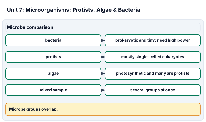
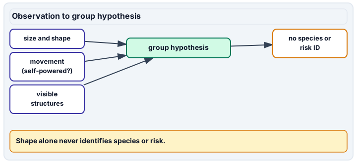
Diagram description
Microbe comparison distinguishes bacteria, protists, algae and mixed samples while acknowledging overlap. Observation to group hypothesis uses size/shape, movement and visible structures to form only a limited group hypothesis.
Data, provenance, and assumptions
| Image set | Source type | Cocci counted | Rods counted | Motile protists tracked |
|---|---|---|---|---|
| Prepared bacteria image | approved supplied image | 18 | 12 | 0 |
| Pond protist image | approved supplied video | 0 | 0 | 6 |
Worked model
In the prepared bacteria image, cocci are 18 of 30 bacterial shapes, or 60%. That is morphology from an approved image, not species identification and not evidence of pathogenicity.
Numerical calibration
- 60 percent cocci among counted bacterial shapes
- 6 tracked motile protists
Attempt the assigned level
Try the tasks before reading the calibration. These are practice answers, not a secure examination; use a new dataset or changed assumption for the assessed transfer.
Foundation: typically grades 7-8
- State the microorganism source boundary in one sentence.
- Calculate the total bacterial shapes counted.
Check after your attempt
- Use approved slides, educator-approved sources or supplied images only; do not culture or amplify unknown microbes.
- 18 cocci + 12 rods = 30 bacterial shapes.
High-school core: typically grades 9-10
- Calculate the cocci percentage and state what it does not prove.
- Explain why "high power" alone is not enough for a bacterial morphology task.
Check after your attempt
- 18/30 = 60% cocci. It does not identify species, pathogenicity or source safety.
- The record needs actual preparation/source, objective/ocular total magnification and resolution; vague high-power language is insufficient.
Honors/readiness extension: quantitative calibration and image-integrity reasoning
- Design a systematic field-sampling plan for approved microorganism images.
- Explain how repeated counts and calibration uncertainty would be reported.
Check after your attempt
- Predefine fields, count the same category rules in each field, keep image/source IDs, avoid duplicate fields, and report counts per field or area.
- Report repeated-count mean/range and the scale or field calibration used; do not hide uncertain or unresolved shapes.
History, reading, and writing connection
History: OpenStax Microbiology 2.3 notes that the relatively rudimentary microscopes of van Leeuwenhoek and his contemporaries were far less powerful than even the most basic microscopes in use today. Use OpenStax Microbiology 2.3 to set a light-microscope boundary: bacteria can be visible at appropriate magnification and preparation, while smaller objects such as viruses are not. Connect that to honest microorganism claims and safe source decisions.
Write in your own words or use an approved accessible equivalent. Cite a specific assigned section or figure, identify its evidence, and state one limitation or counterargument. Use the AI practice contract only for permitted coaching, never to invent observations or write the assessed response.
Transfer to a new case
A student says, "I saw bacteria from my sink at high power." What must be corrected before the claim is acceptable?
Calibration: Reject the improvised unknown source and vague power claim. Use an approved prepared source or supplied image, record objective and ocular total magnification, preparation/stain status, and state that morphology alone does not identify species or risk.
Evidence to retain
Retain approved source, preparation/image type, total magnification, count rules, morphology categories, field IDs and the no-culturing boundary.
Record units, calculations, source/date, uncertainty, and what is measured versus inferred. A simulation or supplied dataset must stay labeled as such. Use only educator-approved slides, specimens, stains, reagents and images. Review SDS information before approved stains, handle slides and coverslips through the approved sharps procedure, and do not culture, amplify or improvise work with unknown microorganisms.
Return to all eight learning pathways. Print this unit page for the student lessons; the linked five-page packet remains the separate assessment companion.
Evidence boundary for this unit
Approved images can assess morphology, counting and limits. Biological handling, prepared-slide use or live tracking are assessed only when the educator observes the approved procedure.
Retain approved source, preparation/image type, total magnification, count rules, morphology categories, field IDs and the no-culturing boundary.
- Science 1: Approved specimen preparation. Observed approved preparation, plus source authorization and no-culturing/no-unknown-microbe statement.
- Science 4: Recognizing bacterial shapes. Total magnification, preparation/image source and morphology-count limit.
- Science 5: Evidence limits & biological handling. Observed handling/disposal route when hands-on work is assigned, plus supplied-image/practical limits.
Course investigation inventory. Completion evidence is a calibrated, honestly documented image or drawing, plus an independently repeatable size or count estimate when the unit calls for one. Supplied-image paper work is distinct from an observed microscope practical.
Unit mastery rubric
| Criterion | Developing | Proficient | Mastery |
|---|---|---|---|
| Approved specimen preparation | Uses unapproved material or cannot follow the selected procedure. | Prepares approved material but loses the field or confuses evidence types. | Follows approved preparation of a risk-assessed slide/specimen (observed), keeps the field and distinguishes live, prepared and recorded evidence; images alone do not credit this criterion; no unknown collection, culturing, enrichment or amplification. |
| Finding & tracking motile protists | Cannot spot a moving organism or loses it the instant it swims. | Sees something move but cannot follow it with the stage. | Locates and tracks a motile protist — amoeba, paramecium, or euglena — steering the stage to keep it in the field. |
| Recognizing algae | Cannot tell algae from other green material. | Suspects algae but cannot point to the feature that marks it. | Recognizes algae by their chloroplasts and form and separates them from plant debris in the mount. |
| Recognizing bacterial shapes | Names features that the image cannot resolve. | Suggests a shape but ignores preparation, scale, or contrast. | Recognizes bacterial morphology only when preparation, total magnification and resolution make it achievable; shape alone never identifies species or pathogenicity. |
| Evidence limits & biological handling | Treats every moving speck as a living organism or ignores source approval. | Uses some distinguishing evidence but overlooks drift or handling limits. | Distinguishes self-powered organisms from drift or debris, states source limits, image/practical distinctions and uncertainty, and follows the approved handling/disposal route whenever hands-on material is used (observed). |
| Integration (cross-domain) | Makes no supported connection between the source and the science. | Uses the source but needs help connecting evidence, writing, or limitations to the science. | Independently connects History, Reading, and Writing using a cited source, appropriate evidence, a limitation, and a scientific explanation. |
Integration is reported separately and cannot lower the science grade or block a science demonstration pass. Science and practical criteria determine that pass. Use the integration guide's evidence checklist for the separately reported criterion.
The approved prepared image was recorded at 1000x total from a 100x objective and 10x ocular. I counted 18 cocci and 12 rods, so cocci were 60%, but I did not identify species or risk.
I used an unapproved source and vague high-power wording, then called anything dot-shaped bacteria without source, scale, or resolution evidence.
Approved images can assess morphology, counting and limits. Biological handling, prepared-slide use or live tracking are assessed only when the educator observes the approved procedure.
A 5-page clipboard packet — unit overview, key terms, the mastery rubric, anchor examples, and a score sheet you can print and grade against.