Unit 08 · The Immune & Integumentary Systems
The year closes with the body’s outer wall and its defense force. This unit covers the skin — its three layers, its jobs from barrier to sensation, how it holds body temperature steady, and how a wound heals — alongside the immune system: the fast, general innate response, the slower, specific adaptive response, the white blood cells and antibodies that carry it out, and how a vaccine trains it. Mastery means you can identify integumentary structures on a model or slide and reason through an immune-response case, exposure to recovery.
Use source-labeled images/models and fictional immune/heat records at the declared level. No wounds, cultures, exposure trials, heat or exercise challenges, patient uploads, or vaccine-history disclosure are requested. Data reasoning stays within the existing science criteria and does not add a new practical.
Student learning: Connect barriers, immune memory, heat transfer, and whole-body limits
Choose the level by readiness, not age alone, and record it before instruction. Foundation, core, and honors tasks are study pathways, not an AP course or a promise of college credit. The instructor retains practical assessment and the published science rubric; integration is reported separately.
Prerequisites: Tissue structure, circulation, cell communication, percentages, and conservation. Core compares response curves; honors considers sampling, heat balance, and passive versus active protection.
Suggested sequence: read and discuss the explanation; attempt the worked model; analyze the data at your selected level; check the answers; then complete the source-linked response and a fresh transfer question. These activities supplement, not replace, supervised practical work and the full-year schedule.
Assigned reading and focus
- OpenStax Anatomy & Physiology 2e, 5.1: Layers of the Skin. Identify epidermis, dermis, and the associated hypodermis on the supplied source figures. Distinguish actual micrographs from diagram labels.
- OpenStax Anatomy & Physiology 2e, 21.2: Barrier Defenses and the Innate Immune Response. Read barriers, phagocytes, signaling, and inflammation. Do not culture microbes, create a wound, or conduct an exposure trial.
- OpenStax Anatomy & Physiology 2e, 21.4: B-lymphocytes and Antibodies. Read B-cell activation, plasma cells, antibodies, and memory. Compare the historical agglutination observation in the introduction with its later molecular interpretation.
Learn the science
Skin proper consists of epidermis and dermis; the hypodermis is associated underlying subcutaneous tissue, not a third layer of skin proper. Epidermis is an avascular keratinized epithelium; dermis contains connective tissue, blood vessels, nerves, and many accessory structures. Hypodermal adipose/connective tissue contributes cushioning, insulation, and attachment. A diagram’s colored boundary does not guarantee the same contrast on a prepared slide.
The barrier reduces water loss and entry of many agents, while receptors support sensation and vessels/glands participate in heat exchange. Sweat cools chiefly when it evaporates, not merely when it appears or drips. Environmental humidity and airflow affect evaporation. Increasing skin blood flow redistributes circulation; maintaining heat transfer and circulating volume interacts with cardiac output, ADH, and renal sodium/water regulation. Our heat table is supplied and must not be reproduced by exercise or heat exposure.
Innate barriers, phagocytes, complement, and inflammatory signaling act rapidly and help activate adaptive responses. Increased local perfusion and permeability recruit cells and proteins, but fluid moving to interstitium can also change effective circulating volume. Inflammation is a coordinated response with possible costs, not a synonym for infection. A micrograph of a white cell alone does not identify a pathogen or establish illness.
Adaptive immunity involves recognition and expansion of lymphocytes with suitable receptors. B cells can differentiate into antibody-secreting plasma cells and memory B cells; T cells coordinate responses or target infected cells, rather than making antibodies themselves. Antigen presentation and helper signals link innate detection and adaptive activation. Antibodies have specificity, but one binding assay is not a complete measure of protection.
Prior exposure to an antigen can support a faster, larger secondary response through memory, though details depend on antigen, timing, and context. Vaccination can establish active immune memory without requiring the natural disease. Passive transfer of antibodies, including maternal IgG across the placenta, can provide temporary support but does not by itself create the recipient’s antigen-specific memory. Neonatal immunity and skin/water regulation develop over time, connecting this unit to Unit 07.
Repair involves overlapping hemostasis, inflammation, tissue formation, and remodeling rather than a rigid stopwatch sequence. A smaller area in a synthetic repair diagram is a geometric observation, not proof that an actual wound is healed, uninfected, or ready for a treatment change. No wounds or photographs of a learner’s skin are requested.
A response ratio needs the same assay scale, a nonzero denominator, and matching comparison times. The arbitrary signal level of 16 below is a classroom comparison threshold, not a protection threshold or a vaccination decision. Sparse sampling gives the first observed crossing, not its exact onset. None of these fictional curves is a clinical antibody result.

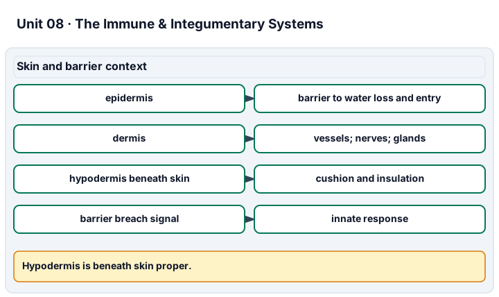
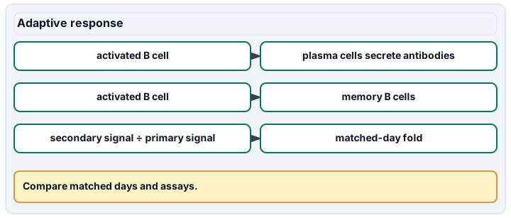
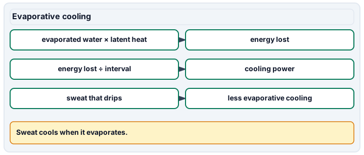
Diagram description
Epidermis is a barrier, dermis contains vessels, nerves and glands, and the hypodermis lies beneath skin proper. An activated B cell can form antibody-secreting plasma cells or memory B cells. Fold comparison uses primary and secondary signals on the same assay day. Evaporative cooling depends on water that evaporates, latent heat, and time.
Data, provenance, and assumptions
| Day after model exposure | Primary signal (arbitrary units) | Secondary signal (arbitrary units) |
|---|---|---|
| 0 | 0 | 4 |
| 4 | 2 | 32 |
| 7 | 8 | 96 |
| 14 | 32 | 128 |
| 21 | 16 | 64 |
| Model | Sweat secreted (g) | Water evaporated (g) | Interval (s) | Latent heat (J/g) |
|---|---|---|---|---|
| A | 600 | 400 | 3600 | 2400 |
| Model day | Diagram area (cm^2) |
|---|---|
| 0 | 9 |
| 4 | 6 |
| 8 | 3 |
Worked model
On day 7, secondary/primary signal = 96/8 = 12. The first sampled values at or above 16 occur on day 14 for primary and day 4 for secondary, a 10-day difference between observed crossings, not exact response onsets. Evaporated fraction = 400/600 × 100 = 66.6667%. Evaporative energy = 400 × 2,400 = 960,000 J, and average power = 960,000/3,600 = 266.6667 W. The diagram area falls by (9 − 3)/9 × 100 = 66.6667%, which alone says nothing about a real injury.
Numerical calibration
- 12 secondary/primary signal
- 10 days between first sampled crossings
- 66.666667 percent
- 266.666667 W
- 66.666667 percent
Attempt the assigned level
Try the tasks before reading the calibration. These are practice answers, not a secure examination; use a new dataset or changed assumption for the assessed transfer.
Foundation
- Identify epidermis, dermis, and hypodermis on a supplied image/model, noting the boundary of skin proper and two visible features.
- Order the broad repair processes and separate barrier, innate, and adaptive examples.
- Compare day-7 signals and calculate the change in the synthetic repair area.
Check after your attempt
- A layered avascular epithelium supports epidermis ID; vascular connective tissue and accessory structures support dermis ID if visible. Hypodermis lies below skin proper.
- Hemostasis, inflammation, tissue formation, remodeling overlap. Skin is a barrier; phagocytes are innate; antigen-specific B/T-cell responses are adaptive.
- The secondary signal is 96 versus 8, or 12 times as large. Area falls by 6 cm^2; this is a diagram measurement, not an injury assessment.
Core
- Compute the day-7 response ratio and first sampled crossings of 16 units. Explain why this is not a clinical protection threshold.
- Compute evaporated fraction and average evaporative power, with unit cancellation.
- Link barrier function, blood flow, renal water conservation, and immune activation in a causal diagram. Do not propose treatment.
Check after your attempt
- Ratio 12; first observed crossings day 14 and day 4, a 10-day difference. Arbitrary assay signal and memory kinetics alone do not define protection or clinical action.
- 66.667% evaporated; energy 960,000 J and average power 266.667 J/s = W. Unevaporated sweat does not contribute that latent-heat loss.
- Skin limits losses and entry, circulation supports transport and heat exchange, and kidneys/hormones regulate fluid handling. Innate signaling recruits defenses and supports adaptive activation; the outcome depends on the integrity and context of all systems.
Honors extension: typically grades 11-12
- Bound when each first threshold crossing could have occurred, assuming continuous increasing signals between the relevant samples. Why can’t the exact lead time be known?
- Predict power if only 200 g evaporates during the same interval. Identify a missing variable before inferring body-temperature change.
- Contrast memory-based secondary responses with maternal passive antibody transfer; explain one reason a single antibody assay cannot summarize immunity.
Check after your attempt
- Primary crossing is after day 7 and no later than day 14; secondary after day 0 and no later than day 4. The 10 days are between sampled crossings; exact crossing times within the intervals remain unknown.
- 200 × 2,400/3,600 = 133.333 W. Metabolic heat, other transfer routes, body heat capacity, and spatial variation would be needed to infer temperature change.
- Active response generates the recipient’s antigen-specific memory; passive antibodies can act temporarily without that memory formation. Cell-mediated defense, antibody specificity/function, and exposure context are not captured by one binding-signal value.
History, reading, and writing connection
Read the historical agglutination account at the start of OpenStax 21.4. Write a cited distinction between the observation (selective clumping) and the later antibody mechanism. Model response: specificity suggests a selective factor, but clumping alone does not reveal its molecular structure or prove protection in every setting. Compare that evidence limit with our arbitrary-unit curves, and explain why ethical consent and non-exposure alternatives matter when studying immunity. Do not recreate the historical exposure or culture work.
Write in your own words or use an approved accessible equivalent. Cite a specific assigned section or figure, identify its evidence, and state one limitation or counterargument. Use the AI practice contract only for permitted coaching, never to invent observations or write the assessed response.
Transfer to a new case
Fresh matched curves have signals 20 and 60 on day 7, but their assay scale is different from the original study. What within-study ratio is justified? Can 60 new units be compared directly to 96 original units or used to decide protection?
Calibration: The within-study ratio is 60/20 = 3. Direct comparison of absolute signals across differently calibrated assays is not justified without a common standard, and neither value establishes a protection decision.
Evidence to retain
Keep source-labeled tissue features, response calculations, interval limits, heat ledger, and a distinction between adaptive memory and passive antibodies. No cultures, injuries, exposure experiments, real patient uploads, or vaccine-history disclosure are part of this evidence.
Record units, calculations, source/date, uncertainty, and what is measured versus inferred. A simulation or supplied dataset must stay labeled as such. Educational fictional cases only: no diagnosis of the learner, real patient uploads, treatment or dosing prescriptions, invasive tests, medical procedure instructions, exercise challenges, breath-holding, or forced personal measurements. No personal, reproductive, or health disclosure is required. Use supplied data and models or an instructor-approved noninvasive demonstration with an agreed alternative. Textbook equations do not certify diagnostic or professional skill; this elective is not a licensed medical course, an AP course, or a claim of college credit.
Return to all eight learning pathways. Print this unit page for the student lessons; the linked five-page packet remains the separate assessment companion.
Unit mastery rubric
| Criterion | Developing | Proficient | Mastery |
|---|---|---|---|
| Skin proper & underlying tissues | Cannot distinguish epidermis, dermis, and hypodermis. | Orders the tissues but needs help with composition or the boundary of skin proper. | Identifies epidermis and dermis as skin proper and hypodermis as underlying tissue, defending their composition/functions using features visible in the assigned image or model. |
| Skin functions & heat balance | Omits barrier/sensory functions or equates sweat secretion with evaporative cooling. | Names the mechanisms but needs help connecting evaporation, blood flow, and fluid regulation. | Explains barrier, sensation, and temperature control; interprets the selected evaporation record with units and links circulation/renal fluid regulation without prescribing care. |
| Repair & evidence limits | Cannot explain the repair processes or mistakes a diagram for clinical evidence. | Orders broad stages but needs help with overlap or interpreting a supplied area change. | Explains overlapping hemostasis, inflammation, tissue formation, and remodeling; interprets the assigned diagram change without inferring healing status from area alone. |
| Innate/adaptive immunity & memory | Confuses phagocytes, B/T cells, antibodies, and memory. | Names defenses but needs help relating activation, vaccination, or passive antibodies. | Distinguishes linked innate/adaptive roles, plasma-cell antibodies and T-cell functions; explains active memory, vaccination, and passive antibody transfer with the selected response-curve evidence. |
| Lab technique (structure ID & immune-case reasoning) | Cannot locate a skin structure on a model or slide, or cannot begin an immune-response case. | Points to a structure or names a defense but cannot connect it to the function or reason through the case. | Locates integumentary structures on a model or slide, defends each structure→function link, and reasons through an immune-response case from exposure to recovery. |
| Integration (cross-domain) | Makes no supported connection between the source and the science. | Uses the source but needs help connecting evidence, writing, or limitations to the science. | Independently connects History, Reading, and Writing using a cited source, appropriate evidence, a limitation, and a scientific explanation. |
Integration is reported separately and cannot lower the science grade or block a science demonstration pass. Science and practical criteria determine that pass. Use the integration guide's evidence checklist for the separately reported criterion.
“Vessels and accessory structures in connective tissue support dermis identification in this image. The hypodermis is below skin proper. In the fictional heat ledger, only 400 of 600 g evaporates: 400 × 2,400 / 3,600 = 266.67 W, not a measured body-temperature change.”
“It’s skin. There are layers. The top one is… the outside? I know sweat comes out somewhere.”
You demonstrate this unit by identifying integumentary structures on a model or a prepared slide — and by reasoning aloud through an immune-response case, from exposure to recovery — not on a multiple-choice test. A criterion counts as mastered only when you can both find the structure at the bench and justify the anatomy and physiology behind it. Mastery is demonstrated, not awarded.
A 5-page clipboard packet — unit overview, key terms, the mastery rubric, anchor examples, and a score sheet you can print and grade against.