What dissection actually teaches.
The squeamish-kid essay. Every parent who calls about the cohort asks some version of this question: my kid isn’t sure they wants to cut a frog open. Good. Neither were any of mine. Here’s what happens between the first incision and the last one.
Here’s the thing nobody warns parents about dissection: the kids who are most worried about it are usually the ones who get the most out of it.
The kids who walk in casual — the ones who’ve been promising for weeks they’re going to “rip it apart” — are almost never the ones holding the scalpel by the second hour. The kids who walked in quiet, gloves a little too tight, are the ones I usually have to drag back to the rest of the schedule because they’ve spent forty minutes on the heart and want to keep going.
That isn’t random. It’s the lesson.
What we’re actually doing at the bench
We dissect a small, finite list of specimens at Bright Minds: an earthworm, a starfish, a clam, a grasshopper, a perch, and the centerpiece, a fetal pig. They are all preserved, ethically sourced from teaching-supply houses that work with the same vendors that supply BSU’s anatomy labs.1 Nothing is killed for our cohort. Every specimen has already entered the educational stream.
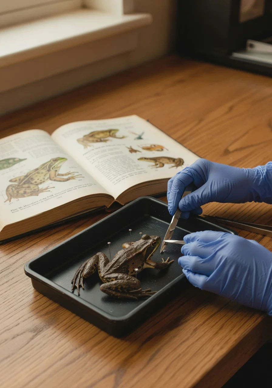
Before any tool comes out of its tray, we do two things. First, the students sketch the specimen, intact, in the lab notebook — name, length, weight, condition. Second, we have a short, honest conversation: this animal lived. We’re going to learn from its body. The fact that it’s small and dead does not make it not worth careful attention.
This is the first thing dissection teaches, and the most important. It is not callousness. It is the opposite. It is reverence. The kids who’ve never done a real dissection sometimes show up expecting it to be a horror movie. They leave understanding that it is closer to a quiet, careful kind of attention — the same attention a surgeon, a veterinarian, or a forensic pathologist would eventually need.
“Reverence is the first thing learned. Callousness, never.”
What a video literally cannot teach
A good dissection video is a useful study tool. I assign them. But three things are not transmissible by screen, and any college anatomy instructor will tell you the same:
Every angle, every neighboring organ, real depth — not a camera’s chosen view.
The textbook shows the canonical case. The bench shows the actual one.
Scalpel depth, scissors over blade — the same family as suturing or drawing blood.
- Three-dimensionality. A video is a 2D projection of a 3D object. The student watching the video sees an organ from one angle, the camera’s. The student at the bench sees it from every angle, in relation to every neighboring organ, with depth and texture and weight. The mental model that gets built is different in kind, not just degree. This is the reason cadaver lab still exists in medical school in the era of CT.2
- Variation. Every specimen is slightly different. The textbook and the video show the canonical case. The bench shows the actual one, with its slightly atypical aortic arch or unusually long mesentery, and the student has to figure out whether the variation matters. That negotiation between the general and the particular — is this normal? is this pathological? am I just looking at it wrong? — is the entire skill of clinical reasoning in a single afternoon.
- Fine motor skill under attention. Holding the scalpel correctly. Knowing the depth of a cut. Choosing the scissors over the scalpel for a delicate membrane. These are motor skills, the same family as suturing, drawing blood, or threading a catheter. They live in the hands and they cannot be downloaded.3
The kid who decides this isn’t for them
Now and then, a student finishes the cohort and decides quietly that medicine, nursing, or research isn’t their path. They found out at fourteen, in a Saturday lab in Boise, instead of at twenty, three semesters into a pre-med program with student loans and a family of expectations.
That is also a win. It might be the biggest one. Career-fit information that arrives this early is one of the most expensive things to come by, and one of the rarest. We’d rather a kid discover, calmly, with no stakes, that the smell of preservative isn’t for them, than have them find out in an OR rotation.
Either they lean in and we’ve given them a head start, or they lean out and we’ve given them a clean answer. Both outcomes are wins. Both are far cheaper to learn at fourteen than at nineteen.
What the parents notice
The phone call I usually get a week or two after the dissection unit isn’t the one parents expect to make. It’s usually some version of: “They came home and explained the circulatory system to their little brother for forty-five minutes. We didn’t know they knew that.”
That’s what dissection actually teaches. Not gore tolerance. Not the appearance of being “advanced.” A genuine, three-dimensional, you-figured-it-out-yourself understanding of how a vertebrate body is put together — and the quiet confidence of someone who knows they have actually seen the thing they’re talking about.
That confidence travels. It shows up in college essays. It shows up in nursing-school interviews. It shows up the first day of gross anatomy, when the kid who dissected a fetal pig at fourteen is the one who’s already comfortable in the room.
We don’t dissect for shock. We dissect because biology is three-dimensional, and the only honest way to learn a three-dimensional thing is in three dimensions, with your hands.
Sources & further reading
- All Bright Minds dissection specimens are preserved teaching specimens sourced from established educational suppliers, including Carolina Biological Supply and equivalents. No animal is killed for cohort use; the same supply chain provides the specimens used in the anatomy labs at Boise State University.
- Ghosh, S. K. (2017). “Cadaveric dissection as an educational tool for anatomical sciences in the 21st century.” Anatomical Sciences Education, 10(3), 286–299. doi:10.1002/ase.1649. A representative review of why hands-on cadaveric work persists in medical education despite the rise of imaging and digital anatomy tools — the three-dimensional variation, the procedural skill, and the professional socialization simply don’t transfer through a screen.
- On procedural and fine-motor skill acquisition as a distinct learning channel from cognitive content learning, see McGaghie, W. C., Issenberg, S. B., Cohen, E. R., Barsuk, J. H., & Wayne, D. B. (2011). “Does simulation-based medical education with deliberate practice yield better results than traditional clinical education? A meta-analytic comparative review of the evidence.” Academic Medicine, 86(6), 706–711. doi:10.1097/ACM.0b013e318217e119. Background on deliberate practice itself: K. Anders Ericsson and Robert Pool, Peak: Secrets from the New Science of Expertise (2016).
Posted Apr 30, 2026.