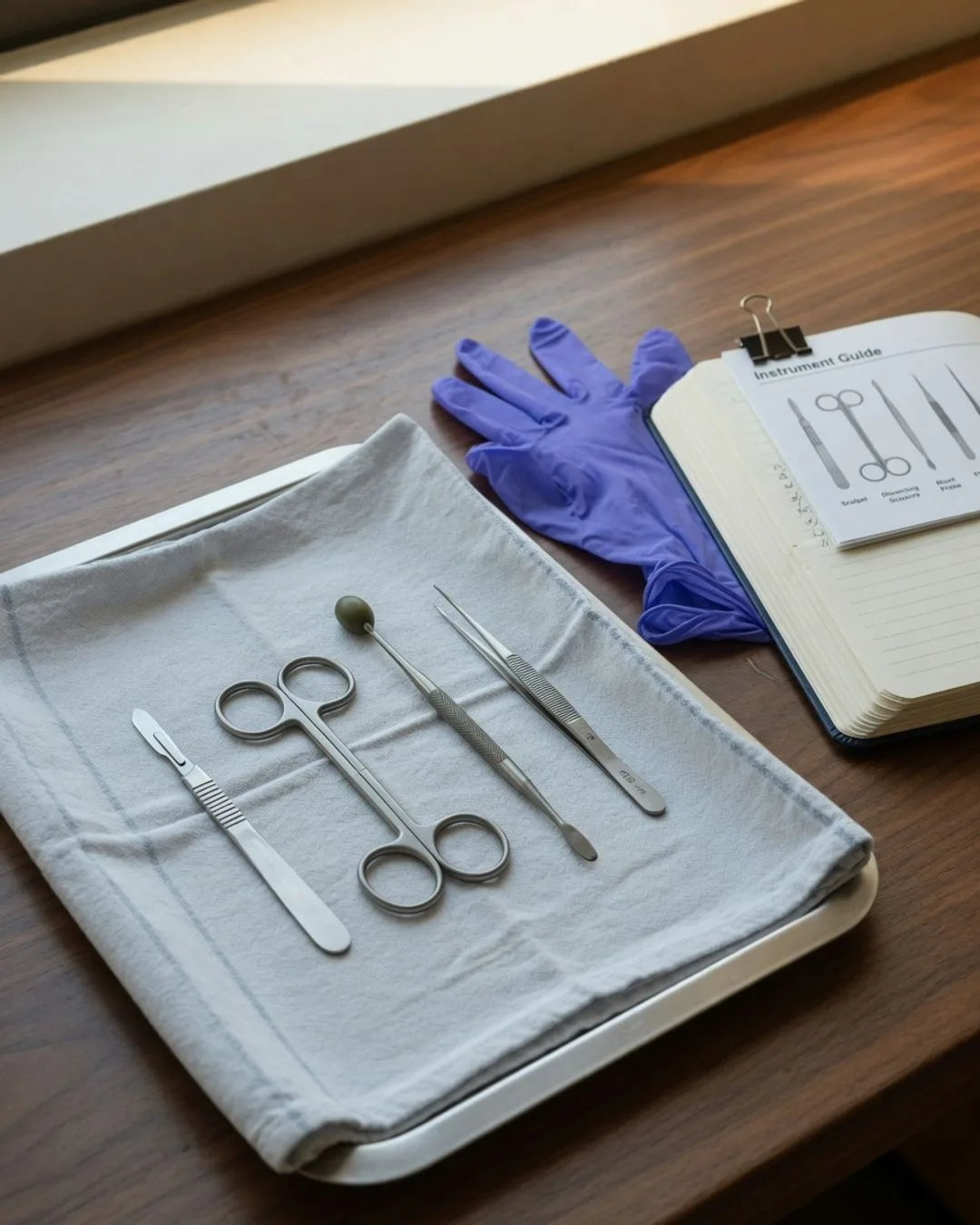
Ask a student who has only studied dissection from a textbook what an earthworm looks like inside, and they will give you a labeled diagram. Ask a student who has actually opened one what it looks like, and they will tell you about the moment the body wall lifted away to reveal the tube nested within a tube, the pale row of hearts, the surprise of how tightly the organs are packed. The first student has a picture someone else drew. The second has a sighting, and the sighting is what the diagram was always trying to stand in for.
That gap is the whole reason this course is built the way it is. Dissection is not a body of facts; it is a set of skills. A diagram, a screen, or a virtual app can only ever show you a cleaned-up, flattened version of an animal that was never flat — the spatial relationships pressed into two dimensions, the variation edited out, the tissue you would have felt reduced to a color on a page. The danger is that the subject collapses into memorizing other people’s drawings: a student learns to label a diagram of a frog without ever once holding a real one and finding the structure themselves.
The bench makes the anatomy real
The job of the dissection is to bring a real, three-dimensional animal up into your own hands — but only for the student willing to look with care. You cannot understand how an organ system fits together from a flat drawing, but you can lift the body wall of an earthworm and see the digestive tract run the whole length of the animal, cradled inside the muscle. You cannot feel the difference between a membrane and the muscle beneath it in a diagram, but you can find it under your own probe and learn that tissue has a texture the page can never show. You cannot be surprised by variation on a screen — but the moment you open a second specimen and it does not quite match the first, you learn something no app could teach: living things vary.
This is what we mean when we say the course is skill-led, not textbook-led. The reading does not come first, with the dissection as a garnish to confirm it. The bench comes first. The observation is made where it actually lives (specimen in the tray, probe in hand, handled with care from the first cut), and the textbook is the tool we reach for to name what we just exposed. A student who has traced a nerve along a real body is ready to be told what it does. A student who has only been told about it is ready to forget it.
The diagram in the book is a claim about something real. The bench is where the student finds out the claim is true — by exposing it themselves.
What the bench teaches that the page cannot
Beyond making anatomy concrete, the bench teaches a set of things a textbook structurally cannot, because they are not facts — they are skills and judgments that only form under real conditions:
- That careful observation is hard. Deciding whether the pale line under your probe is a nerve or a strand of connective tissue, telling a real structure from something your own cut created, waiting to look before you move — these teach a patience that no labeled drawing ever will.
- That real specimens don’t look like the diagram. The organ sits a little differently than the textbook drew it. A structure hides behind another. Two animals of the same species are not built quite the same. Learning to reason about why the real specimen departs from the clean drawing is the heart of the skill.
- That the cut serves the look. How you open the body wall, how deep you go, how you pin it back, how gently you separate a layer — the answer depends on the doing, and the doing can only be learned by doing. The cut is never the point; it is only there to let you see, and it is made with respect for the animal that made the lesson possible.
The two-day rhythm
Practically, this conviction becomes a schedule. The course runs on a two-day rhythm. One day is the Concept Day: the anatomy and the technique are introduced up close — where a structure sits, how an incision is made, how a system is laid open. The next is the Experiment Day, where that same technique becomes physical at the bench and gets written into a real lab notebook in the student’s own hand. Between the two days, the student studies at home, and that gap is not dead time. It is where the diagram and the specimen knit together into something that lasts.
We are not against the textbook; a serious dissection course needs a rigorous one, and this course has it. We are against the textbook going first and the bench going second, because we have watched what that produces: a student who can label every part of a diagram and has never once found one of those parts on a real animal with their own hands. Put the bench first, and dissection stops being a labeled drawing. It becomes a thing the student has actually seen and done, which is the only kind of dissection anyone remembers.