Unit 02 · The Earthworm
This unit uses earthworm segmentation as direct anatomical evidence while avoiding rank language. A preserved specimen is one individual with artifacts, not a universal body.
Student learning: The Earthworm
Choose the route and level by prerequisites, access needs, and approved supervision before instruction. Foundation, core, and honors tasks are study pathways for this Grades 8-12 semester lab-skills course, not an AP course, college credit, or credential claim.
Prerequisites: Can orient a bilaterally symmetric specimen and record a measurement with units.
Suggested sequence: read the assigned section, study the teaching explanation, analyze the labeled synthetic data, attempt the selected level, check the answer, then complete a fresh transfer defense. These lessons supplement the assessment packet and do not authorize any procedure.
Assigned reading and focus
- OpenStax Biology 2e: Animal Form and Function. [earthworm-segmentation] Use the animal body-plan discussion to connect bilateral symmetry and body organization to the earthworm specimen.
- OpenStax Biology 2e: Organizing Life on Earth. [earthworm-segmentation] Use the phylogenetic-tree explanation to avoid treating an earthworm as a step toward a later animal.
- Darwin Online: Vegetable Mould and Worms. Use Darwin’s earthworm work as the history anchor: careful observation of a small animal can support a large soil-process claim without ranking animals.
Learn the science
Choose readiness by prerequisites and route approval, not age alone. Public worked answers are nonsecure practice, and no page authorizes a procedure outside the educator-approved plan.
Earthworm segmentation without ranking animals. The earthworm unit teaches segmentation, orientation, and internal organization. Segmentation is real anatomical evidence, but it is not a rank on a ladder; it is one body-plan pattern in a branching animal history.
Assumptions: The synthetic specimens have intact anterior-posterior orientation and visible external segment rings, but preservation may stretch, curl, flatten, or obscure internal landmarks.
Uncertainty: Segment counts vary with specimen length, preservation, and how incomplete terminal rings are counted. The numbers are practice measurements, not a population study.

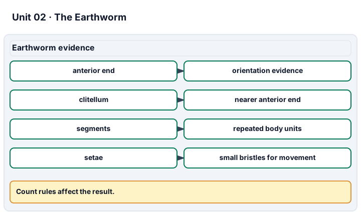
Diagram description
The grid uses anterior end, clitellum, segments, and setae as evidence. Segment density uses total visible segments divided by total length from the supplied records. Incomplete terminal rings and preservation artifacts are stated in the description rather than folded into the formula.
Data, provenance, and assumptions
| Specimen | Length (cm) | Visible segments | Preservation note |
|---|---|---|---|
| E1 | 9.6 | 95 | slightly curled |
| E2 | 10.2 | 102 | posterior tip flattened |
Paper investigation sequence and evidence record
Scope and safety: No improvised procedures, preservatives, latex or other allergens, or instruments are introduced by these public lessons. Hands-on work must follow the educator-approved procedure and SDS, use ethically and legally sourced preserved specimens, and respect opt-out access to an equivalent image-based, virtual, or model route without penalty. No live-animal procedures are part of this course.
Materials and preparation
- [earthworm-segmentation] Use the supplied earthworm-segments records, assigned source sections, calculator and an accessible graph or diagram. No physical apparatus is required.
Procedure and schedule
- [earthworm-segmentation] Question: How does repeated segmentation help orient and identify the earthworm specimen? Prerequisites: Can orient a bilaterally symmetric specimen and record a measurement with units.
- [earthworm-segmentation] Variables: Specimen length, visible segment count, orientation, and preservation condition. Controls: Count only visible rings under the approved procedure; do not infer missing rings from memory. Replication: Repeat the count after rotating the tray and compare with a partner or image-based route.
- [earthworm-segmentation] Analysis: Compute visible segments per centimeter and describe why it is only local practice evidence. Review: An educator reviews prerequisites, source interpretation, reasoning and accessibility before assessment. A paper response does not certify unobserved practical technique; approved hands-on work requires a separate record.
Record: Retain earthworm-segmentation, the selected level, original inputs and first attempt, source/date, system and axes, units, assumptions, calculation/graph, uncertainty, correction and fresh transfer. Link only to the stated science criteria.
Worked model
The two synthetic worms have 95 + 102 = 197 visible segments across 9.6 + 10.2 = 19.8 cm, giving about 9.95 segments per cm.
Numerical calibration
- 9.9494949495 visible segments per cm
Attempt the assigned level
Try the tasks before reading the calibration. These are practice answers, not a secure examination; use a new specimen, image set, model, or supervised task for assessment.
Foundation: guided specimen literacy and source-backed vocabulary
- Label anterior, posterior, dorsal, ventral, and one repeated segment on the synthetic record.
Check after your attempt
- The answer must use directional terms and identify repetition without saying the earthworm is a lesser version of another animal.
High-school core: typically grades 9-10, semester lab-skills course with independent evidence and approved routes
- Explain how segmentation supports an earthworm identification while naming one preservation artifact that can mislead the count.
Check after your attempt
- Repeated rings and internal organization support the identification; curling, flattening, or damaged ends can change the count and must be recorded.
Honors extension: typically grades 11-12, quantitative morphology and comparative physiology
- Compare segment density across the two specimens and explain why the result is morphology practice, not a universal earthworm body value.
Check after your attempt
- The combined density is about 9.95 segments/cm. It is a practice summary from two preserved specimens and cannot define all earthworms or infer ancestry by itself.
History, reading, and writing connection
Use Darwin’s earthworm work, OpenStax phylogeny, and animal form to explain segmentation on a branching tree. State what the earthworm specimen shows and what phylogenetic evidence would add.
Write in your own words or use an approved accessible equivalent. Cite a specific assigned section, identify its evidence, and state one limitation.
Transfer to a new case
A third worm has 88 visible segments over 8.9 cm. Is that enough to call it the same species as E1 and E2?
Calibration: No. 88/8.9 is about 9.89 segments/cm, similar to the practice records, but species identity needs additional morphology and source evidence.
Evidence to retain
Completion evidence: the student defends a real specimen identification, explains a valid cross-specimen comparison, and states the limits of the evidence. The student must identify a real earthworm structure and explain what segmentation does and does not prove.
Record route, units, source/date, observed structures, calculations, comparison category, uncertainty, and what is measured versus inferred. No improvised procedures, preservatives, latex or other allergens, or instruments are introduced by these public lessons. Hands-on work must follow the educator-approved procedure and SDS, use ethically and legally sourced preserved specimens, and respect opt-out access to an equivalent image-based, virtual, or model route without penalty. No live-animal procedures are part of this course.
Return to all eight learning pathways. Print this unit page for student learning; the linked five-page packet remains the separate assessment companion.
Evidence, routes, and comparison limits
Integration is reported separately and cannot lower the science grade or block a science demonstration pass. Science and practical criteria determine that pass.
Completion evidence: the student defends a real specimen identification, explains a valid cross-specimen comparison, and states the limits of the evidence. The student must identify a real earthworm structure and explain what segmentation does and does not prove.
Equivalent routes: Image-based, virtual, model, and professionally supervised routes can meet the same conceptual standard without penalty. Practical skill is credited only when hands-on technique is observed on an approved route.
Safety and ethics: No improvised procedures, preservatives, latex or other allergens, or instruments are introduced by these public lessons. Hands-on work must follow the educator-approved procedure and SDS, use ethically and legally sourced preserved specimens, and respect opt-out access to an equivalent image-based, virtual, or model route without penalty. No live-animal procedures are part of this course.
- Science 1: Instrument handling & safe technique. Only an educator-observed approved practical or professionally supervised route can evidence Instrument handling & safe technique; supplied data, images, virtual routes, or models can support planning and explanation but cannot credit hands-on skill. Rubric row.
- Science 2: Careful exposure & observation. Only an educator-observed approved practical or professionally supervised route can evidence Careful exposure & observation; supplied data, images, virtual routes, or models can support planning and explanation but cannot credit hands-on skill. Rubric row.
- Science 4: Locating & naming internal structures. Supplied images and instructor-selected diagrams can evidence internal-structure naming only for visible or labeled organs; actual exposure quality requires observed practical work. Rubric row.
- Science 5: Explaining structure & function (and specimen care). The segment-density calculation, artifact note, and branching-tree explanation evidence structure-function reasoning and specimen-care limits. Rubric row.
Unit mastery rubric
| Criterion | Developing | Proficient | Mastery |
|---|---|---|---|
| Instrument handling & safe technique | Grips the scalpel, scissors, forceps, or probe awkwardly; cuts too hard or too deep; puts hands or specimen at risk. | Holds the instruments correctly with reminders and cuts more carefully, but still presses too hard or steadies the specimen poorly. | Holds each instrument the right way and makes shallow, controlled cuts, working safely for both the student and the specimen; this technique criterion requires educator-observed approved practical work. |
| Careful exposure & observation | Cuts before looking and tears or destroys structures while opening the specimen. | Opens the specimen roughly along the correct line but disturbs organs before observing them. | Opens the specimen cleanly along the dorsal midline without damaging structures, and observes the layout before disturbing anything; this exposure criterion requires educator-observed approved practical work. |
| Locating & naming external structures | Cannot point to the clitellum, segments, or setae, or tell anterior from posterior. | Finds a few external landmarks with prompting but confuses dorsal and ventral or miscounts segments. | Locates and names the clitellum, segments, and setae on the specimen, approved image, model, or virtual route and orients it correctly by anterior/posterior and dorsal/ventral. |
| Locating & naming internal structures | Guesses at the organs or names the wrong ones once the specimen is open. | Finds the larger organs but cannot reliably distinguish crop from gizzard or trace the blood vessel and nerve cord. | Locates and names the dorsal blood vessel, aortic arches, crop, gizzard, intestine, seminal vesicles, nephridia, and ventral nerve cord on the approved specimen, image, model, or virtual route. |
| Explaining structure & function (and specimen care) | Cannot say what a structure does, and lets the specimen dry out or handles it carelessly. | Explains one or two structures' functions but not the rest, and keeps the specimen moist only when reminded. | Explains why key structures do their jobs while stating segment-count variation, preservation artifacts, and route limits; specimen-care technique is credited only when observed. |
| Integration (cross-domain) | Makes no supported connection between the source and the science. | Uses the source but needs help connecting evidence, writing, or limitations to the science. | Independently connects History, Reading, and Writing using a cited source, appropriate evidence, a limitation, and a scientific explanation. |
Integration is reported separately and cannot lower the science grade or block a science demonstration pass. Science and practical criteria determine that pass. Use the integration guide's evidence checklist for the separately reported criterion.
“This pale band is the clitellum — it’s segments 32 to 37, and it secretes the cocoon. The dark line down the dorsal side is the dorsal blood vessel; I traced it forward to the five pairs of aortic arches that pump the blood. Just behind them the crop stores food and the gizzard grinds it, which is why the gizzard wall feels muscular.”
“I think that tube is the stomach? I cut it before I really looked, so it’s kind of torn now. I’m not sure which end is the front.”
You demonstrate this unit by doing the dissection while an instructor watches — locating, exposing, and naming real structures and explaining their function on your own specimen, not a written test. Technique criteria are mastered only when an educator observes them; identification and explanation criteria can be mastered on the approved specimen, image, model, or virtual route without penalty. Mastery is demonstrated, not awarded.
A 5-page clipboard packet — unit overview, key terms, the mastery rubric, anchor examples, and a score sheet you can print and grade against.