Unit 03 · The Cardiovascular System
This unit follows the blood: the four heart chambers and the valves that keep it moving one way, the cardiac cycle you can hear as “lub-dub,” the split between the pulmonary and systemic circuits, the arteries, veins, and capillaries that carry it, and what blood pressure and pulse actually measure. Mastery means you can take a blood-pressure and pulse reading, interpret it, and trace a drop of blood through the whole loop — not just label a diagram.
Declare either supplied pressure/pulse/sound records with a model defense, or separately approved noninvasive equipment work with an available record alternative. No personal measurements, diagnoses, or medical procedures are required. Record interpretation does not certify cuff or stethoscope technique; selected calculations belong to these criteria, not a new practical.
Student learning: Reason from cardiac volumes to circulation and oxygen delivery
Choose the level by readiness, not age alone, and record it before instruction. Foundation, core, and honors tasks are study pathways, not an AP course or a promise of college credit. The instructor retains practical assessment and the published science rubric; integration is reported separately.
Prerequisites: Anatomical directions, muscle contraction, volume units, and ratios. Honors uses steady-state mass balance and an explicitly simplified pressure model.
Suggested sequence: read and discuss the explanation; attempt the worked model; analyze the data at your selected level; check the answers; then complete the source-linked response and a fresh transfer question. These activities supplement, not replace, supervised practical work and the full-year schedule.
Assigned reading and focus
- OpenStax Anatomy & Physiology 2e, 19.4: Cardiac Physiology. Read Resting Cardiac Output and the factors affecting stroke volume. Use the equations and supplied fictional values only; do not follow exercise examples or interpret personal readings.
- OpenStax Anatomy & Physiology 2e, 20.2: Blood Flow, Blood Pressure, and Resistance. Read Variables Affecting Blood Flow and A Mathematical Approach. Compare pressure difference, flow, resistance, and the assumptions in Poiseuille’s model.
- OpenStax Anatomy & Physiology 2e, 22.4: Gas Exchange. Read gas partial-pressure gradients and the ventilation/perfusion discussion. Link a lung receiving air to a lung receiving blood; a pressure is not an oxygen-content measurement.
Learn the science
Trace systemic veins → right atrium → tricuspid valve → right ventricle → pulmonary valve → pulmonary arteries → lung capillaries → pulmonary veins → left atrium → mitral valve → left ventricle → aortic valve → systemic arteries and capillaries. Artery and vein describe direction relative to the heart, not oxygenation. Both circuits carry red blood. At steady state their time-averaged flows match; do not add the two ventricular outputs as if they were separate deliveries to the body.
Valves open and close because of pressure differences, not because they actively pump. Ventricular filling, isovolumetric contraction, ejection, and isovolumetric relaxation form a useful cycle model. S1 is associated chiefly with atrioventricular valve closure; S2 with semilunar closure. A supplied sound recording and cycle diagram can be aligned, but listening to a learner’s chest is not required.
Stroke volume is end-diastolic minus end-systolic ventricular volume. Ejection fraction is stroke volume divided by end-diastolic volume, usually expressed as a percentage. Cardiac output equals heart rate times stroke volume. Heart rate alone does not give cardiac output: filling time, preload, afterload, and contractility can change the volume ejected per beat.
Arteries have walls suited to higher pressure, veins are more compliant reservoirs, and capillaries provide a thin exchange interface. Systolic and diastolic pressures are not flow rates. Mean arterial pressure ≈ diastolic + one third of pulse pressure is a resting-cycle approximation, not an exact average for every waveform or heart rate. Effective systemic resistance = (mean arterial pressure − right-atrial pressure)/cardiac output in this lumped, steady-state model.
Plasma transports dissolved substances and proteins; red cells carry hemoglobin; white cells participate in defense; platelets participate in hemostasis. Oxygen delivery requires blood flow and oxygen content, not saturation or pulse alone. For the supplied contents, delivery = output × arterial content; steady-state tissue oxygen use = output × (arterial − mixed-venous content). Oxygen bound to hemoglobin makes content different from partial pressure.
The kidneys link long-term extracellular volume regulation to venous return and pressure, while baroreflexes and autonomic signals act quickly. These relationships are conditional, not a rule that more retained water always solves low output. A single fictional dataset cannot identify the cause of a changed circulation, diagnose illness, or authorize treatment.

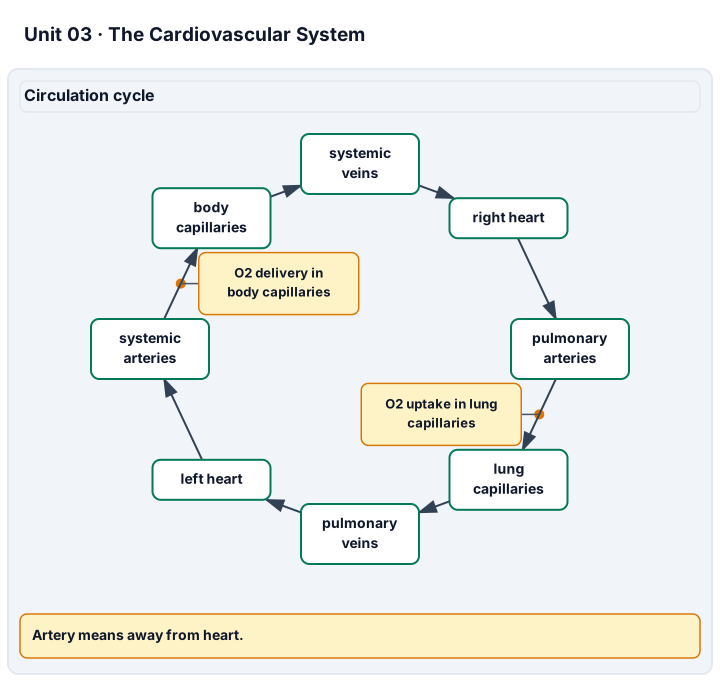
Diagram description
Blood returns by systemic veins to the right heart, travels through pulmonary arteries to lung capillaries, returns by pulmonary veins to the left heart, and leaves through systemic arteries to body capillaries. Artery and vein refer to direction relative to the heart, not oxygenation. Calculations include stroke volume, ejection fraction, cardiac output, oxygen delivery, and tissue oxygen use from supplied records.
Data, provenance, and assumptions
| Record | Heart rate (beats/min) | End-diastolic volume (mL) | End-systolic volume (mL) |
|---|---|---|---|
| A | 75 | 120 | 50 |
| B | 100 | 110 | 60 |
| Record | Systolic pressure (mmHg) | Diastolic pressure (mmHg) | Right-atrial pressure (mmHg) |
|---|---|---|---|
| A | 120 | 80 | 5 |
| Record | Arterial O2 content (mL O2/L blood) | Mixed-venous O2 content (mL O2/L blood) |
|---|---|---|
| A | 200 | 150 |
Worked model
For A, SV = 120 − 50 = 70 mL/beat; EF = 70/120 × 100 = 58.3333%; output = 75 × 70 = 5,250 mL/min = 5.25 L/min. Delivery is 5.25 × 200 = 1,050 mL O2/min, whereas tissue use is 5.25 × (200 − 150) = 262.5 mL O2/min. Estimated mean pressure is 80 + 40/3 = 93.3333 mmHg and effective resistance is (93.3333 − 5)/5.25 = 16.8254 mmHg min/L. These are distinct quantities with distinct denominators.
Numerical calibration
- 70 mL/beat
- 58.333333 percent
- 5.25 L/min
- 262.5 mL O2/min
- 1050 mL O2/min
- 93.333333 mmHg
- 16.825397 mmHg min/L
Attempt the assigned level
Try the tasks before reading the calibration. These are practice answers, not a secure examination; use a new dataset or changed assumption for the assessed transfer.
Foundation
- Trace the two circuits and locate all four valves on a model; explain why a pulmonary artery is still an artery.
- Subtract the supplied volumes to find stroke volume in A and B. Which record ejects more per beat?
- Match plasma, red cells, white cells, and platelets to transport, gas carriage, defense, and hemostasis.
Check after your attempt
- A pulmonary artery carries blood away from the right ventricle toward the lungs. The right and left sides connect the pulmonary and systemic circuits in series.
- A ejects 70 mL/beat and B ejects 50 mL/beat; A ejects more per beat.
- Plasma transports dissolved substances and proteins; red cells carry hemoglobin, white cells perform immune roles, and platelets support hemostasis. These roles interact rather than operating as isolated boxes.
Core
- Calculate output and ejection fraction for both records. Test the claim “a faster heart rate must mean a greater output.”
- Calculate oxygen delivery and oxygen use for A; explain why they differ.
- Align supplied S1/S2 labels with valve closure in a cycle sketch, and distinguish pulse pressure from mean pressure.
Check after your attempt
- A: 5.25 L/min and 58.33%; B: 5.00 L/min and 45.45%. B has the higher rate but lower output, so rate alone is insufficient.
- Delivery is 1,050 mL O2/min and use is 262.5 mL O2/min. Venous blood retains oxygen; tissues do not extract every delivered molecule.
- S1 corresponds chiefly to AV-valve closure and S2 to semilunar closure. Pulse pressure is 40 mmHg for A; the model mean pressure is about 93.33 mmHg, not (120 + 80)/2.
Honors extension: typically grades 11-12
- Compute effective systemic resistance including right-atrial pressure. State the steady-state and waveform assumptions.
- For a rigid tube with fixed pressure difference, length, and viscosity, predict the flow ratio if radius becomes 0.8 of its initial value.
- Could normal-looking oxygen saturation guarantee A’s oxygen delivery? Explain what else would be needed.
Check after your attempt
- About 16.8254 mmHg min/L, assuming the supplied mean-pressure approximation and steady-state average flow. Pulsatile elastic vessels are not a single rigid resistor.
- Poiseuille’s ideal laminar model gives 0.8^4 = 0.4096 of initial flow. A network with active regulation, changing pressure, or turbulence need not follow this isolated-tube prediction.
- No. Hemoglobin amount, arterial content, and output also matter; saturation is a fraction of binding capacity, not total oxygen content or tissue delivery.
History, reading, and writing connection
OpenStax 20.2 identifies Poiseuille and presents his flow relationship. Cite that section and compare an ideal rigid-tube explanation with the living circulation. Model response: the fourth-power radius term gives a testable prediction under fixed conditions, but compliant, branching, actively regulated vessels limit its direct application. Connect this use of quantitative evidence to the older circulation question without claiming that our synthetic records are Harvey’s measurements.
Write in your own words or use an approved accessible equivalent. Cite a specific assigned section or figure, identify its evidence, and state one limitation or counterargument. Use the AI practice contract only for permitted coaching, never to invent observations or write the assessed response.
Transfer to a new case
A new fictional ventricle has HR 80 beats/min, EDV 130 mL, ESV 65 mL, and arterial-minus-venous O2 content 40 mL/L. Find output and oxygen use. Can those numbers alone name a disease?
Calibration: SV = 65 mL/beat, output = 5.2 L/min, and modeled oxygen use = 208 mL O2/min at steady state. No: causes, measurement uncertainty, and clinical context are not provided, and this is not a diagnostic exercise.
Evidence to retain
Retain the two-circuit sketch, cycle explanation, calculations, units, and unknown-variable list. A supplied pressure or sound record does not establish cuff, stethoscope, or patient-examination proficiency.
Record units, calculations, source/date, uncertainty, and what is measured versus inferred. A simulation or supplied dataset must stay labeled as such. Educational fictional cases only: no diagnosis of the learner, real patient uploads, treatment or dosing prescriptions, invasive tests, medical procedure instructions, exercise challenges, breath-holding, or forced personal measurements. No personal, reproductive, or health disclosure is required. Use supplied data and models or an instructor-approved noninvasive demonstration with an agreed alternative. Textbook equations do not certify diagnostic or professional skill; this elective is not a licensed medical course, an AP course, or a claim of college credit.
Return to all eight learning pathways. Print this unit page for the student lessons; the linked five-page packet remains the separate assessment companion.
Unit mastery rubric
| Criterion | Developing | Proficient | Mastery |
|---|---|---|---|
| Heart chambers & valves | Cannot name the four chambers or the valves between them. | Names the four chambers but cannot say which valve guards each or which side pumps where. | Names the four chambers and the valves on a model or specimen, and explains which side pumps to the lungs and which to the body. |
| Cardiac cycle, sounds & output | Cannot connect filling/ejection, valves, and stroke volume. | Describes the cycle but needs help interpreting supplied volumes or sound labels. | Links the cycle and S1/S2 to valve closure; calculates stroke volume, ejection fraction, and output from supplied inputs at the declared level, not from pulse alone. |
| Systemic & pulmonary circulation & blood vessels | Cannot tell the two circuits apart or name the three vessel types. | Names arteries, veins, and capillaries but cannot trace blood through the pulmonary and systemic loops. | Distinguishes arteries, veins, and capillaries by structure and traces a drop of blood through both the pulmonary and systemic circuits. |
| Pressure, blood composition & oxygen transport | Confuses pressure, flow, or blood-component roles. | Names the quantities and components but needs help explaining their relationships. | Explains pulse and systolic/diastolic pressure, plasma/cell/platelet roles, and the selected oxygen-content or resistance calculation with units and steady-state assumptions. |
| Pressure, pulse & sound evidence | Cannot read the assigned record or carry out the declared approved setup. | Reads values but needs help connecting them to anatomy, units, or limitations. | Under the declared record or approved equipment mode, interprets pressure, pulse, and sound evidence with units and anatomical reasoning; distinguishes recorded from inferred quantities without diagnosing. |
| Integration (cross-domain) | Makes no supported connection between the source and the science. | Uses the source but needs help connecting evidence, writing, or limitations to the science. | Independently connects History, Reading, and Writing using a cited source, appropriate evidence, a limitation, and a scientific explanation. |
Integration is reported separately and cannot lower the science grade or block a science demonstration pass. Science and practical criteria determine that pass. Use the integration guide's evidence checklist for the separately reported criterion.
“The supplied cycle places S1 at AV-valve closure and S2 at semilunar closure. Record A ejects 120 − 50 = 70 mL per beat; at 75 beats/min that is 5.25 L/min. A pulse alone would not supply stroke volume or oxygen content.”
“The heart goes lub-dub. Blood pressure is… a number? I felt something at the wrist but I don’t know what it was.”
Defend the declared model/record task and trace blood through both circuits. If a separately approved noninvasive equipment pathway is chosen, its technique is observed and documented separately from record analysis; an equivalent supplied-record pathway is available before instruction. Do not claim a measurement or clinical skill that was not performed.
A 5-page clipboard packet — unit overview, key terms, the mastery rubric, anchor examples, and a score sheet you can print and grade against.