Unit 04 · The Respiratory System
Breathing looks simple until you measure it. This unit follows air from the airways through the bronchial tree to the alveoli, the muscles and pressure changes that move it in and out, the gas exchange that loads oxygen and unloads carbon dioxide at the alveolar wall, and the lung volumes you can put a number on with a spirometer. Mastery means you can measure your own lung volumes and defend what gas exchange is actually doing, not just label a diagram.
The declared default is a supplied spirogram and airway-model defense. Any equipment demonstration needs separate instructor approval and an agreed record alternative. No forced breathing, breath-holding, exercise challenge, or personal lung-volume measurement is required. Honors acid-base practice is not another required practical or a medical qualification.
Student learning: Separate ventilation, gas exchange, and bounded acid-base responses
Choose the level by readiness, not age alone, and record it before instruction. Foundation, core, and honors tasks are study pathways, not an AP course or a promise of college credit. The instructor retains practical assessment and the published science rubric; integration is reported separately.
Prerequisites: Gas diffusion, pressure, blood flow, and volume/time units. Core uses subtraction and multiplication; honors uses base-10 logarithms and model limits.
Suggested sequence: read and discuss the explanation; attempt the worked model; analyze the data at your selected level; check the answers; then complete the source-linked response and a fresh transfer question. These activities supplement, not replace, supervised practical work and the full-year schedule.
Assigned reading and focus
- OpenStax Anatomy & Physiology 2e, 22.3: The Process of Breathing. Read pressure relationships, volumes/capacities, and respiratory control. Analyze supplied records only; do not copy forced-breathing or exercise examples.
- OpenStax Anatomy & Physiology 2e, 22.4: Gas Exchange. Read partial pressure, gas solubility, and ventilation/perfusion. Distinguish dissolved-gas pressure from total oxygen content.
- OpenStax Anatomy & Physiology 2e, 26.4: Acid-Base Balance. Read buffers and respiratory/renal regulation, focusing on time scales and mechanisms. Disease tables are not a learner-screening tool.
Learn the science
The conducting airways carry and condition air from trachea through bronchi to smaller bronchioles; alveoli provide a thin exchange surface. Cartilage supports larger airways, while smooth muscle helps regulate smaller-airway resistance. During quiet inspiration, respiratory muscle action expands the thorax and lowers alveolar pressure relative to atmospheric pressure. Quiet expiration is largely passive recoil; an unqualified “lungs suck in oxygen” misses the pressure mechanism.
Minute ventilation is breathing frequency × tidal volume. Some air remains in conducting space without exchanging with blood on that breath. In the simplified model, alveolar ventilation = frequency × (tidal volume − dead-space volume). Equal minute ventilation can therefore yield different alveolar ventilation. The supplied dead space is an assumption, not something to estimate from a classmate.
Vital capacity = inspiratory reserve + tidal volume + expiratory reserve. Residual volume remains after maximal expiration; ordinary spirometry cannot measure residual volume directly, so it also cannot independently give total lung capacity. A separate supplied residual-volume estimate can be added. Reading a fictional spirogram is not performing spirometry, and there is no breath-holding, hyperventilation, or personal lung-capacity challenge here.
Oxygen diffuses down its own partial-pressure gradient from alveoli toward incoming capillary blood; carbon dioxide follows its own gradient in the opposite direction. Diffusion depends on barrier thickness, surface area, and gas properties as well as the gradient. Ventilation brings air and perfusion brings blood: an air-filled region with little perfusion cannot make its usual contribution to oxygenating circulating blood.
Much oxygen is carried bound to hemoglobin, whereas carbon dioxide is transported mainly as bicarbonate, with dissolved and protein-bound fractions. CO2 + H2O ⇌ H2CO3 ⇌ H+ + HCO3− links gas exchange to pH. Buffers limit a change but do not remove an unlimited acid load. Carbon dioxide excretion changes the volatile-acid component; renal hydrogen-ion excretion and bicarbonate handling address different parts of the balance.
Use the supplied teaching equation pH = 6.1 + log10([HCO3−]/(0.03 × PCO2)). Bicarbonate is in mmol/L, PCO2 in mmHg, and the CO2 solubility coefficient is 0.03 mmol/(L mmHg), at an approximately 37 °C model condition. The argument of the logarithm is dimensionless. The apparent pK and coefficient are approximations; the equation does not by itself identify a cause or a treatment.
Chemical buffering is rapid, respiratory responses can act over minutes, and renal adaptation takes hours to days. Compensation need not restore the original pH or remove the primary disturbance. In our deliberately bounded example, bicarbonate falls from 24 to 12 mmol/L and PCO2 may fall only as far as a stipulated 30 mmHg floor. This floor is invented for the problem, not a clinical compensation rule or a safe breathing target. Full restoration of the reference 20:1 ratio would need 20 mmHg, outside that model’s permitted range.

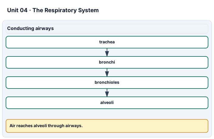
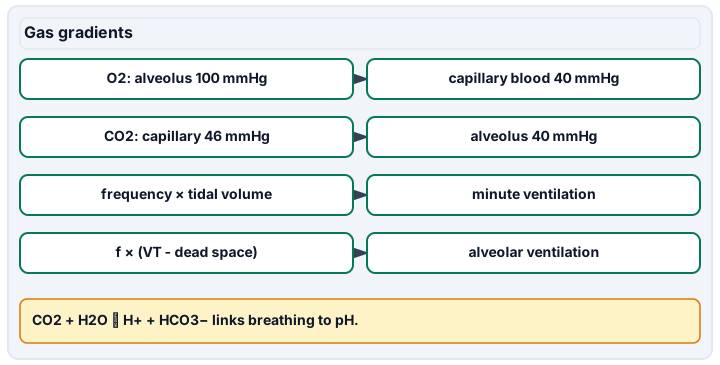
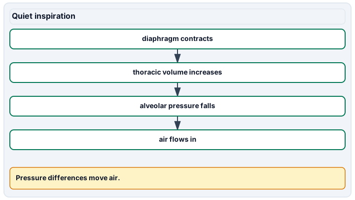
Diagram description
Air flows through conducting airways to alveoli. Oxygen moves from alveoli toward incoming blood down its partial-pressure gradient; carbon dioxide moves in the opposite direction. Minute ventilation and alveolar ventilation use different formulas. Quiet inspiration follows muscle contraction, larger thoracic volume, lower alveolar pressure, and inward air flow.
Data, provenance, and assumptions
| Pattern | Frequency (breaths/min) | Tidal volume (L/breath) | Dead space (L/breath) |
|---|---|---|---|
| A | 12 | 0.5 | 0.15 |
| B | 20 | 0.3 | 0.15 |
| Component | Volume (L) |
|---|---|
| Inspiratory reserve | 3 |
| Tidal | 0.5 |
| Expiratory reserve | 1.1 |
| Residual: separate estimate | 1.2 |
| Gas | Alveolar partial pressure (mmHg) | Incoming pulmonary-capillary partial pressure (mmHg) |
|---|---|---|
| Oxygen | 100 | 40 |
| Carbon dioxide | 40 | 46 |
| State | Bicarbonate (mmol/L) | PCO2 (mmHg) |
|---|---|---|
| Reference | 24 | 40 |
| Reduced bicarbonate, unchanged CO2 | 12 | 40 |
| Bounded respiratory response | 12 | 30 |
| Raised CO2, unchanged bicarbonate | 24 | 60 |
| Illustrative later renal response | 30 | 60 |
Worked model
Both A and B move 6 L/min in total, but A delivers 12 × (0.5 − 0.15) = 4.2 L/min of alveolar ventilation and B only 3 L/min. Vital capacity is 3 + 0.5 + 1.1 = 4.6 L; adding the separate 1.2 L residual estimate gives 5.8 L total capacity. In the bicarbonate model the reference ratio is 24/(0.03 × 40) = 20 and pH ≈ 7.40103. Halving bicarbonate at fixed CO2 gives pH 7.1; allowing PCO2 to fall to 30 gives pH 7.224939, only partial recovery. With raised PCO2 of 60 and later bicarbonate 30, pH is 7.321849, still not the reference.
Numerical calibration
- 6 L/min
- 4.2 L/min
- 3 L/min
- 4.6 L
- 5.8 L
- 7.40103 pH (dimensionless)
- 7.1 pH (dimensionless)
- 7.224939 pH (dimensionless)
- 7.321849 pH (dimensionless)
- 20 mmHg
Attempt the assigned level
Try the tasks before reading the calibration. These are practice answers, not a secure examination; use a new dataset or changed assumption for the assessed transfer.
Foundation
- Trace air to alveoli and explain how expanding the thorax changes airflow direction.
- Calculate minute ventilation in both patterns and vital capacity from the first three volume entries.
- Use the gas-pressure table to draw oxygen and carbon-dioxide diffusion arrows. State why no personal measurements are needed.
Check after your attempt
- Trachea → bronchi → bronchioles → alveoli. Expansion lowers alveolar pressure below atmospheric pressure, so air flows inward; the airway path and muscle action are different parts of the explanation.
- A and B each have 6 L/min minute ventilation. Vital capacity is 4.6 L. These are supplied model records, not a demonstration of equipment technique.
- O2 moves from 100 toward 40 mmHg, while CO2 moves from 46 toward 40 mmHg. All necessary inputs are fictional and supplied.
Core
- Calculate alveolar ventilation in A and B and total capacity using the separately supplied residual volume.
- Explain how poor perfusion can limit gas exchange even when ventilation is present. Connect this to cardiac output in Unit 03.
- Compare pH direction for reduced bicarbonate at fixed CO2 and for raised CO2 at fixed bicarbonate; name which organ system can influence each variable.
Check after your attempt
- Alveolar ventilation: A 4.2 L/min, B 3.0 L/min. Total capacity: 5.8 L. Ordinary spirometry alone does not supply the residual value.
- Gas transfer requires both air and blood; cardiac output and its regional distribution affect perfusion. Total ventilation is not identical to oxygen delivery.
- Both changes lower the ratio and pH. Lungs regulate CO2 removal; kidneys reclaim filtered bicarbonate and generate new bicarbonate while excreting acid equivalents. These processes have different time scales.
Honors extension: typically grades 11-12
- Recompute every pH in the table, retaining units until the logarithm. Explain why the bounded respiratory response cannot restore the reference ratio.
- Assume unchanged CO2 production and that alveolar PCO2 is inversely proportional to alveolar ventilation at steady state. If A corresponds to 40 mmHg, predict B’s value and state two limitations.
- Explain why an unchanged pH can conceal two changed variables, rather than proving normal physiology.
Check after your attempt
- Reference 7.40103; reduced bicarbonate 7.10000; bounded response 7.224939; raised CO2 7.224939; later renal response 7.321849. Restoring a 20:1 ratio with bicarbonate 12 requires PCO2 20 mmHg, below the stipulated 30 floor.
- 40 × 4.2/3.0 = 56 mmHg in the ideal steady-state model. Changing CO2 production, dead space, uneven ventilation/perfusion, or lack of equilibration defeats this prediction; it is not a breathing instruction.
- Bicarbonate and CO2 can change proportionally and preserve their ratio. A ratio alone cannot establish whether either input or its cause is unchanged.
History, reading, and writing connection
Cite the pressure-volume explanation in OpenStax 22.3 and the regulation time scales in 26.4. Write a response explaining why a successful equation is not a complete account of a person. Model response: Boyle’s relationship isolates a physical condition, while a breathing organism exchanges gases, changes muscle activity, and regulates renal chemistry. State which assumptions our synthetic records hold fixed and one kind of evidence a real physiological research claim would additionally need.
Write in your own words or use an approved accessible equivalent. Cite a specific assigned section or figure, identify its evidence, and state one limitation or counterargument. Use the AI practice contract only for permitted coaching, never to invent observations or write the assessed response.
Transfer to a new case
A new model has bicarbonate 18 mmol/L and PCO2 30 mmHg. Find its ratio and pH. Does matching the reference pH show that neither variable changed?
Calibration: 18/(0.03 × 30) = 20, so pH ≈ 7.40103. Both variables are lower than the reference values, despite the same ratio. No diagnostic or treatment conclusion follows.
Evidence to retain
Keep the pressure arrows, volume decomposition, dead-space comparison, unit-bearing pH calculations, and a compensation-limit statement. Do not collect breathing traces, force an expiration, or label the modeled response as a performed respiratory test.
Record units, calculations, source/date, uncertainty, and what is measured versus inferred. A simulation or supplied dataset must stay labeled as such. Educational fictional cases only: no diagnosis of the learner, real patient uploads, treatment or dosing prescriptions, invasive tests, medical procedure instructions, exercise challenges, breath-holding, or forced personal measurements. No personal, reproductive, or health disclosure is required. Use supplied data and models or an instructor-approved noninvasive demonstration with an agreed alternative. Textbook equations do not certify diagnostic or professional skill; this elective is not a licensed medical course, an AP course, or a claim of college credit.
Return to all eight learning pathways. Print this unit page for the student lessons; the linked five-page packet remains the separate assessment companion.
Unit mastery rubric
| Criterion | Developing | Proficient | Mastery |
|---|---|---|---|
| Airways & the bronchial tree | Cannot name the major airways or order them from the trachea to the alveoli. | Names the airways but cannot trace the path air takes or say where cartilage gives way to smooth muscle. | Traces air from the trachea through bronchi and bronchioles to the alveoli on a model, and explains how each airway's structure fits its job. |
| Mechanics of breathing | Describes breathing as air being “sucked in” with no reference to muscles or pressure. | Names the diaphragm but cannot connect its movement to the pressure change that drives airflow. | Explains how the diaphragm and rib cage change thoracic volume and pressure, and predicts airflow direction from that pressure change. |
| Gas exchange, transport & acid-base balance | Confuses gas gradients, carriage, or the role of carbon dioxide in pH. | Explains one mechanism but needs help linking perfusion, gas carriage, and buffering. | Explains partial-pressure diffusion, ventilation/perfusion, and gas carriage; connects CO2/bicarbonate to pH at the selected level, with explicit limits on respiratory and renal compensation. |
| Lung volumes, ventilation & control | Confuses tidal/vital/residual volume or total/alveolar ventilation. | Reads volumes but needs help with dead space, residual-volume limits, or control. | Interprets supplied tidal/reserve volumes and capacities, distinguishes total from alveolar ventilation, and explains CO2-related control; states that ordinary spirometry does not measure residual volume. |
| Spirogram & model evidence | Cannot read the assigned trace or identify its axes. | Reads volumes but needs help separating supplied estimates from measured trace values. | Defends the declared spirogram/model or approved demonstration evidence, reads volumes with units, and identifies uncertainty and separately supplied values without claiming unperformed spirometry. |
| Integration (cross-domain) | Makes no supported connection between the source and the science. | Uses the source but needs help connecting evidence, writing, or limitations to the science. | Independently connects History, Reading, and Writing using a cited source, appropriate evidence, a limitation, and a scientific explanation. |
Integration is reported separately and cannot lower the science grade or block a science demonstration pass. Science and practical criteria determine that pass. Use the integration guide's evidence checklist for the separately reported criterion.
“The supplied volumes give vital capacity 4.6 L; residual volume is a separate estimate, not measured by ordinary spirometry. Both patterns move 6 L/min, but their alveolar ventilation is 4.2 versus 3.0 L/min because dead-space air must be subtracted.”
“You breathe in air and breathe out air. The lungs are for oxygen. Vital capacity is… how much you can breathe?”
Read the provided trace, defend airway structure and gas exchange, and distinguish assumptions from observations. The published alternative assesses interpretation, not personal lung function or medical equipment competence. Any separately authorized demonstration must have an equivalent non-personal option and is not authorized by these reading pages.
A 5-page clipboard packet — unit overview, key terms, the mastery rubric, anchor examples, and a score sheet you can print and grade against.