Unit 05 · The Nervous System & Senses
The nervous system is the body's signaling network, and you can watch it work. This unit builds from the single neuron and the electrochemical signal that races down its axon, across the synapse to the next cell, out to the divisions of the brain and spinal cord, and into the reflex arcs and special senses you can test on a partner. Mastery means you can run a reflex or sensory test and trace the signal through the arc, not just recite the parts of a neuron.
Declare a diagram/record pathway or a separately approved noninvasive demonstration with a record alternative. No electrical stimulation, partner reflex examination, or personal response measurement is required. Supplied timing analysis does not certify test administration, and leveled practice is not a new required practical.
Student learning: Distinguish neuronal mechanisms, reflex control, and response-time evidence
Choose the level by readiness, not age alone, and record it before instruction. Foundation, core, and honors tasks are study pathways, not an AP course or a promise of college credit. The instructor retains practical assessment and the published science rubric; integration is reported separately.
Prerequisites: Membrane transport, electrical sign conventions, distance/time units, and the difference between a stimulus and an observation. Honors uses logarithms and bounded measurement uncertainty.
Suggested sequence: read and discuss the explanation; attempt the worked model; analyze the data at your selected level; check the answers; then complete the source-linked response and a fresh transfer question. These activities supplement, not replace, supervised practical work and the full-year schedule.
Assigned reading and focus
- OpenStax Anatomy & Physiology 2e, 12.4: The Action Potential. Read membrane potential, channel gating, refractory periods, and conduction. Use the supplied diagrams and traces rather than any electrical stimulation.
- OpenStax Anatomy & Physiology 2e, 15.2: Autonomic Reflexes and Homeostasis. Read the somatic/visceral reflex comparison and baroreceptor pathway. Omit clinical examination boxes; identify sensors, integration, and effectors.
Learn the science
Dendrites and the cell body receive and integrate many inputs; an axon carries action potentials toward terminals. The CNS is brain and spinal cord; the PNS includes nerves and ganglia outside them. Cerebral, cerebellar, brainstem, and spinal circuits make different contributions, but a simple region-to-job label is not a complete map of thought or behavior.
Electrochemical gradients and selective channel permeability produce membrane voltage. Sodium-potassium ATPase maintains concentration gradients over time; it does not directly cause each fast upstroke. In a typical neuronal action potential, voltage-gated sodium entry drives depolarization and potassium conductance contributes to repolarization. Threshold, channel inactivation, and refractory periods constrain firing. Stronger stimuli can change firing patterns rather than making every action potential proportionally taller.
At a chemical synapse, a presynaptic action potential opens calcium channels; calcium supports transmitter release, and transmitter binds receptors on the postsynaptic cell. Receptor and channel properties determine excitation or inhibition. This organization usually gives the synapse a functional direction, but electrical synapses also exist; “all neurons have a chemical gap that can only work one way” is too broad.
A somatic reflex links receptor → sensory pathway → CNS integration → motor pathway → muscle. Some reflex pathways integrate in the spinal cord, but ascending information and descending brain modulation still exist. Special senses use specialized receptors: photoreceptors for light, cochlear hair cells for sound, vestibular hair cells for balance, and receptor cells for taste and smell. Detecting a stimulus, deciding on a response, and executing movement all take time.
Baroreceptors sense vessel stretch and help brainstem circuits adjust autonomic output to the heart and vessels. Osmoreceptors inform hypothalamic regulation of ADH; renal sodium/water handling changes the controlled system more slowly. Receptors detect a variable, not the name of a disease, and neither pathway is demonstrated by asking a learner to produce an abnormal response.
Whole reaction time is not nerve conduction time. A separately supplied two-site record permits a segment estimate only if the downstream synaptic and muscle delays are common and cancel. Speed = distance/(proximal latency − distal latency). Keep milliseconds and seconds distinct. Timing uncertainty broadens the range, and the calculation does not authorize electrical tests or reflex testing on a partner.
For honors, E_K = 61.5 × log10([K+]outside/[K+]inside) mV is a supplied single-ion equilibrium approximation for a monovalent ion near 37 °C. It is not the actual resting membrane potential of a multi-ion cell. Raising extracellular potassium makes this equilibrium value less negative; predicting excitability additionally needs sodium-channel availability and other conductances. A renal electrolyte change can influence neurons and muscle without a one-number diagnostic rule.

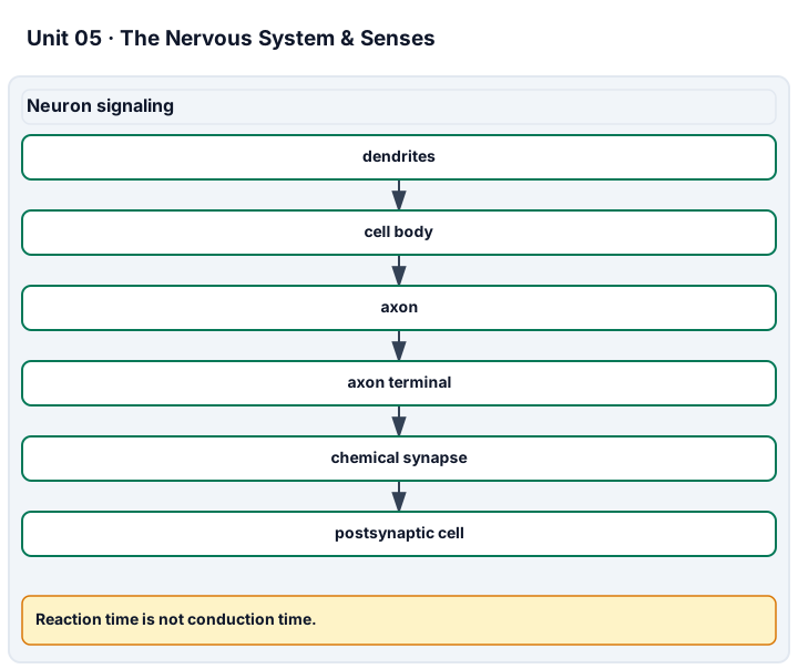
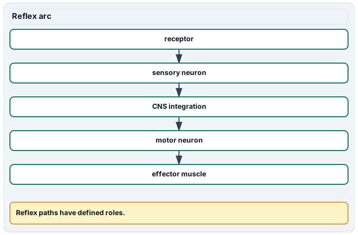
Diagram description
Dendrites, cell body, axon, axon terminal, chemical synapse, and postsynaptic cell describe one signalling path. A reflex arc is separate: receptor, sensory neuron, CNS integration, motor neuron, effector muscle. Conduction speed uses distance and latency difference converted from milliseconds to seconds, not whole reaction time.
Data, provenance, and assumptions
| Record | Segment length (m) | Proximal latency (ms) | Distal latency (ms) | Each latency uncertainty (ms) |
|---|---|---|---|---|
| Segment A | 0.24 | 8 | 4 | 0.2 |
| Trial | Whole response time (ms) |
|---|---|
| R1 | 220 |
| R2 | 240 |
| R3 | 260 |
| R4 | 280 |
| State | Outside K+ (mmol/L) | Inside K+ (mmol/L) |
|---|---|---|
| A | 4 | 140 |
| B | 8 | 140 |
Worked model
The latency difference is 8 − 4 = 4 ms = 0.004 s; speed = 0.24/0.004 = 60 m/s. Worst-case difference bounds are 3.6 and 4.4 ms, giving speeds from 54.5455 to 66.6667 m/s; do not average the latency errors away without a model of them. Whole reaction times average 250 ms and cannot substitute for the 4 ms segment delay. The supplied equilibrium equation gives E_K,A = −94.960185 mV and E_K,B = −76.446840 mV, a +18.513345 mV shift.
Numerical calibration
- 60 m/s
- 54.545455 m/s
- 66.666667 m/s
- 250 ms
- -94.960185 mV
- 18.513345 mV
Attempt the assigned level
Try the tasks before reading the calibration. These are practice answers, not a secure examination; use a new dataset or changed assumption for the assessed transfer.
Foundation
- Label a neuron and trace a reflex pathway on supplied diagrams, including the CNS integration site.
- Match vision, hearing, and balance to their receptor types. Explain why an observed response includes more than axon conduction.
- Find mean and range of the four whole response times.
Check after your attempt
- Afferent signals travel toward CNS integration; efferent signals leave toward an effector. A spinal integration site does not mean the brain receives no information or cannot modulate the response.
- Photoreceptors detect light; cochlear and vestibular hair cells support hearing and balance. Sensory transduction, integration, decision, and motor execution contribute to a whole response.
- Mean = 250 ms; range = 280 − 220 = 60 ms. Four trials from one session are not a population estimate.
Core
- Calculate segment speed and explain why subtracting the latencies, rather than using either whole latency, is appropriate under the stated conditions.
- Explain the channel sequence in an action potential and the different role of the sodium-potassium pump.
- Build a baroreflex sensor-to-effector diagram and compare its time scale with renal volume regulation.
Check after your attempt
- 0.24 m/0.004 s = 60 m/s. Subtraction removes the stipulated common downstream delay; if that delay differs, the calculation no longer isolates the segment.
- Sodium-channel activation, inactivation, and potassium conductance shape the spike; the ATPase maintains the concentration gradients. Pumping and channel flow are not interchangeable processes.
- Vessel stretch → sensory afferents → brainstem integration → altered autonomic output → heart/vessel response. Renal sodium/water regulation contributes over a longer interval; neither is a clinical test here.
Honors extension: typically grades 11-12
- Find the worst-case speed interval and name an omitted uncertainty.
- Recompute E_K in both states and the shift. Explain why a potassium value alone cannot predict an action-potential trace.
- Could a group difference in reaction time prove a change in nerve conduction? Propose additional supplied evidence instead of a personal test.
Check after your attempt
- About 54.545–66.667 m/s if distance is exact. Distance measurement error and failure of common-delay cancellation would add uncertainty.
- −94.960185 and −76.446840 mV; change +18.513345 mV. Multiple ions, channel states, and membrane properties determine a real trace.
- No. Task familiarity, attention, sensory processing, and motor delay can differ. An independently characterized conduction dataset with matching conditions would test a narrower claim.
History, reading, and writing connection
Cite OpenStax 12.4’s action-potential graph and 15.2’s reflex diagram. Write an evidence critique of “a slower response proves slower nerves.” Model response: a voltage trace and a behavioral timing record measure different events; a mechanism requires evidence that separates component delays. Explain why the development of time-resolved electrical recording added evidence that anatomical drawings alone could not supply. Do not invent an experiment or quotation for Cajal or Galvani.
Write in your own words or use an approved accessible equivalent. Cite a specific assigned section or figure, identify its evidence, and state one limitation or counterargument. Use the AI practice contract only for permitted coaching, never to invent observations or write the assessed response.
Transfer to a new case
A fresh supplied segment is 0.18 m long and the latency difference is 3 ms. A separate response time is 300 ms. Compute segment speed and explain which number cannot be used as its denominator.
Calibration: 0.18/0.003 = 60 m/s. The 300 ms response includes other processes and is not the segment’s conduction time.
Evidence to retain
Retain the annotated circuit, latency subtraction, uncertainty interval, and a clear distinction between the hypothetical equilibrium potential and a measured trace. No sensory, electrical, or reflex examination of a person is required.
Record units, calculations, source/date, uncertainty, and what is measured versus inferred. A simulation or supplied dataset must stay labeled as such. Educational fictional cases only: no diagnosis of the learner, real patient uploads, treatment or dosing prescriptions, invasive tests, medical procedure instructions, exercise challenges, breath-holding, or forced personal measurements. No personal, reproductive, or health disclosure is required. Use supplied data and models or an instructor-approved noninvasive demonstration with an agreed alternative. Textbook equations do not certify diagnostic or professional skill; this elective is not a licensed medical course, an AP course, or a claim of college credit.
Return to all eight learning pathways. Print this unit page for the student lessons; the linked five-page packet remains the separate assessment companion.
Unit mastery rubric
| Criterion | Developing | Proficient | Mastery |
|---|---|---|---|
| Neuron structure & electrical signaling | Confuses neuronal parts or ion-channel and pump roles. | Labels the neuron but needs help with gradients, channel sequence, or refractory behavior. | Labels the neuron and explains ion-channel action potentials and gradient maintenance; interprets the selected electrical model without equating a single-ion equilibrium value with a whole-cell voltage. |
| Synapses & neurotransmission | Cannot distinguish a chemical synapse from axonal conduction. | Names transmitter release but needs help linking calcium, receptors, and response. | Explains calcium-dependent release and receptor response at a chemical synapse, its usual functional direction, and why electrical synapses prevent a universal one-way-gap claim. |
| CNS & PNS: brain and spinal cord | Cannot separate the central from the peripheral nervous system or locate the major brain regions. | Divides CNS from PNS but confuses the major brain regions or their jobs. | Distinguishes CNS from PNS, locates the major brain regions and the spinal cord on a model, and states what each region does. |
| Reflexes, senses & homeostatic control | Cannot trace a sensor-to-effector pathway or identify special-sense receptors. | Names pathway parts but needs help ordering them or explaining modulation. | Traces receptor, sensory input, integration, motor output, and effector; links special senses to receptors and compares autonomic feedback with spinal integration and brain modulation. |
| Reflex & timing evidence | Cannot interpret the assigned response record or pathway. | Reads a timing value but confuses whole response with segment conduction or omits units. | Defends the declared record/model or approved demonstration, distinguishes reaction from conduction time, and uses the supplied distance, latency differences, and uncertainty at the agreed level. |
| Integration (cross-domain) | Makes no supported connection between the source and the science. | Uses the source but needs help connecting evidence, writing, or limitations to the science. | Independently connects History, Reading, and Writing using a cited source, appropriate evidence, a limitation, and a scientific explanation. |
Integration is reported separately and cannot lower the science grade or block a science demonstration pass. Science and practical criteria determine that pass. Use the integration guide's evidence checklist for the separately reported criterion.
“The diagram shows spinal integration, not a pathway isolated from brain modulation. In the supplied timing record, subtracting common downstream delays gives 4 ms for 0.24 m, or 60 m/s. The 250 ms average whole response is a different quantity.”
“You hit the knee and the leg kicks. Nerves send signals to the brain. A neuron is a brain cell, I think.”
Defend the selected pathway and the provenance of its supplied timing record. No partner examination is needed. If a separate approved demonstration is selected, document exactly the noninvasive task and alternative beforehand; analysis of synthetic latencies does not claim that a conduction test was performed.
A 5-page clipboard packet — unit overview, key terms, the mastery rubric, anchor examples, and a score sheet you can print and grade against.