Unit 02 · Cells & Their Structures
Every living thing is made of cells — the tiny building blocks of life. In this unit you’ll learn the cell theory, compare plant and animal cells, and find out what each part does, from the cell membrane to the nucleus to the mitochondria. Best of all, you’ll use a real microscope to see cells for yourself. Mastery means you can spot cell parts under the lens and explain the job each one does.
Student learning: What can a microscope image support?
Choose the level by readiness, not age alone, and record it before instruction. Foundation, core, and honors tasks are study pathways, not an AP course or a promise of college credit. The instructor retains practical assessment and the published science rubric; integration is reported separately.
Prerequisites: Cell versus organism, ratios, millimetres and micrometres; distinguish a schematic from a micrograph.
Suggested sequence: read and discuss the explanation; attempt the worked model; analyze the data at your selected level; check the answers; then complete the source-linked response and a fresh transfer question. These activities supplement, not replace, supervised practical work and the full-year schedule.
Assigned reading and focus
- OpenStax Biology 2e, 4.1: Studying Cells. Read Microscopy, Light Microscopes, and Cell Theory; compare Figures 4.2 and 4.3. Distinguish Hooke's 1665 cork observations, Leeuwenhoek's 1670s microorganisms, and the later development of cell theory. Checked 2026-09-27.
- OpenStax Biology 2e, 4.3: Eukaryotic Cells. Study Figure 4.8 and the sections The Plasma Membrane, The Cytoplasm, The Nucleus, Mitochondria, and Plant Cells. Use the diagram to learn functions, not as proof that every labeled part is visible with a school microscope. Checked 2026-09-27.
Learn the science
Cells exchange materials through a membrane. Cytoplasm supports reactions; the nucleus in most eukaryotic cells contains DNA; mitochondria support energy transfer through cellular respiration. Photosynthetic plant tissues have chloroplasts, but not every plant cell does. A plant cell wall is distinct from its membrane.
Hooke named the compartments he saw in cork in 1665; Leeuwenhoek described microorganisms in the 1670s. Neither one image nor one observer established all of modern cell theory. The later work of Schleiden, Schwann, Virchow, and others connected evidence across tissues and organisms.
Magnification enlarges an image; resolution separates adjacent features. A classroom microscope cannot reliably identify every organelle in a general cell diagram. Label an observed outline or visible chloroplast only when the actual image supports it; write not resolved for unsupported features.
Use a scale bar that belongs to the image. Cell length = image cell length / image scale-bar length x the scale-bar value. Resizing the entire image changes both displayed lengths equally and leaves the estimated real size unchanged.

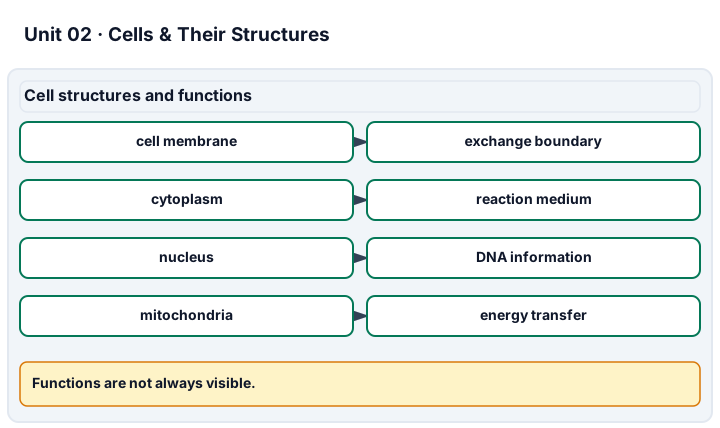
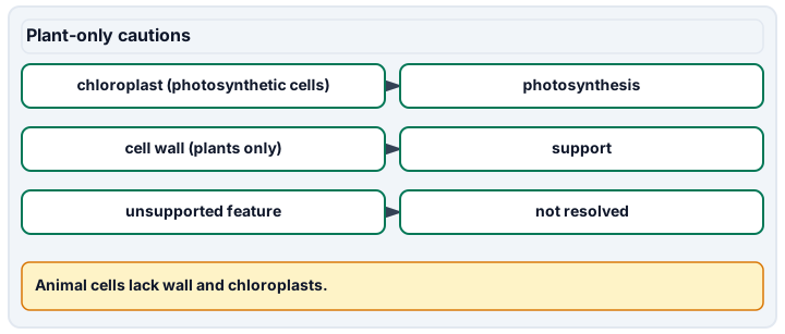
Diagram description
Cell membrane, cytoplasm, nucleus, and mitochondria are paired with functions. Chloroplasts are limited to photosynthetic cells, and cell walls are plant structures not found in animal cells. Unsupported features from a microscope image should be marked not resolved. Scale-bar calculations use the source scale only.
Data, provenance, and assumptions
| Image | Scale-bar value (micrometres) | Displayed bar (mm) | Displayed cell (mm) |
|---|---|---|---|
| A | 50 | 20 | 40 |
| B | 50 | 40 | 80 |
| Eyepiece power | Objective power |
|---|---|
| 10 | 40 |
Worked model
For A, 40/20 x 50 = 100 micrometres. For B, 80/40 x 50 = 100 micrometres again. The specified lenses give 10 x 40 = 400 times magnification. Neither number proves that a mitochondrion was visible.
Numerical calibration
- 100 micrometres in either supplied image record
- 400 times
Attempt the assigned level
Try the tasks before reading the calibration. These are practice answers, not a secure examination; use a new dataset or changed assumption for the assessed transfer.
Foundation: typically grades 7-8
- Annotate your own simple plant/animal cell diagrams using Figure 4.8: membrane, cytoplasm, nucleus, mitochondrion, plant wall, and a chloroplast in a photosynthetic cell. Add a function to each.
- Submit a two-column Hooke/Leeuwenhoek timeline and distinguish a reference-diagram label from an observation in a micrograph.
Check after your attempt
- Membrane: exchange boundary; cytoplasm: reaction medium; nucleus: DNA; mitochondrion: cellular respiration; wall: support; chloroplast: photosynthesis. Animals lack cell walls and chloroplasts.
- 1665 cork compartments and 1670s microorganisms are different evidence. A drawn label is not evidence that you personally resolved that structure.
Core
- Calculate cell size for A and B and the lens magnification with units. Explain why resizing does not double the biological cell.
- Use the assigned light/electron micrographs to list one supported feature and one unresolved feature; identify image method and preparation as limitations.
Check after your attempt
- Both lengths are 100 micrometres; magnification is 400 times. The numerator and denominator resize together.
- A defensible answer identifies features actually visible in the selected figure and its caption; electron-microscope detail is not automatically available in a live light-microscope view.
Extension
- If A's cell length could be 38-42 mm while its scale bar is 20 mm, calculate the possible size range.
- Explain why "I zoomed in, therefore I discovered a new organelle" confuses evidence with display size.
Check after your attempt
- 38/20 x 50 to 42/20 x 50 gives 95-105 micrometres under the stated fixed-bar assumption.
- Digital enlargement does not add resolved detail; source resolution, preparation, contrast, and alternative interpretations must be examined.
History, reading, and writing connection
Original response: Prepare a dated evidence note comparing Hooke's cork observation, Leeuwenhoek's microorganisms, and modern cell theory using OpenStax 4.1. Add your scale calculation and one captioned feature from an assigned micrograph. State explicitly whether each item is a historical report, supplied model, or your separately recorded observation; finish with one instrument limit. A letter from Leeuwenhoek is not required or silently assumed to be supplied.
Write in your own words or use an approved accessible equivalent. Cite a specific assigned section or figure, identify its evidence, and state one limitation or counterargument. Use the AI practice contract only for permitted coaching, never to invent observations or write the assessed response.
Transfer to a new case
A different image has a 10 mm bar labeled 20 micrometres and a cell 15 mm long. Is this cell 400 micrometres long because the microscope says 400 times?
Calibration: No. 15/10 x 20 = 30 micrometres. Magnification is not a length, and a copied image may have been resized.
Evidence to retain
Cells & cell theory: dated evidence note and three cell-theory statements. Plant vs. animal cells: annotated comparison with the photosynthetic-tissue qualification. Cell parts & their jobs: labels with functions. Cells as living systems: explain exchange and energy processing together. Lab technique (using a microscope): separately observed safe focusing of an approved prepared slide and a labeled, scaled record; supplied measurements do not demonstrate handling. Retain calculations, figure/caption references, uncertainty, transfer, and the separately reported integration note.
Record units, calculations, source/date, uncertainty, and what is measured versus inferred. A simulation or supplied dataset must stay labeled as such. This is reading, paper-model, and data work, not authorization to collect pond water, swab people, culture organisms, dissect, expose wildlife, or ingest study materials. Use only instructor-approved prepared slides, images, or in-room materials for separately observed practical skills. Supplied data are not your observations.
Return to all eight learning pathways. Print this unit page for the student lessons; the linked five-page packet remains the separate assessment companion.
Unit mastery rubric
| Criterion | Developing | Proficient | Mastery |
|---|---|---|---|
| Cells & cell theory | Thinks only some living things, like animals, are made of cells. | Says living things are made of cells but can’t explain the cell theory. | Explains that all living things are made of cells and states the cell theory in their own words. |
| Plant vs. animal cells | Can’t tell a plant cell from an animal cell. | Names differences but assumes every plant cell has chloroplasts. | Compares plant and animal cells, distinguishing the cell wall from the membrane and chloroplasts in photosynthetic tissues. |
| Cell parts & their jobs | Can name one or two parts but not what they do. | Lists the parts but matches only some to their jobs. | Names the main parts — cell membrane, nucleus, cytoplasm, mitochondria, and more — and explains the job of each. |
| Cells as living systems | Thinks a cell is just a blob with no working parts. | Knows cells are busy but can’t connect a part to a life process. | Explains how the parts work together so the cell can get energy, grow, and stay alive. |
| Lab technique (using a microscope) | Cannot yet handle or focus an approved prepared slide safely. | Gets an image but needs help focusing, scaling or distinguishing visible features from inferred ones. | Safely focuses an approved prepared slide at two powers, records scale and visible features, and identifies unresolved structures. |
| Integration (cross-domain) | Makes no supported connection between the source and the science. | Uses the source but needs help connecting evidence, writing, or limitations to the science. | Independently connects History, Reading, and Writing using a cited source, appropriate evidence, a limitation, and a scientific explanation. |
Integration is reported separately and cannot lower the science grade or block a science demonstration pass. Science and practical criteria determine that pass. Use the integration guide's evidence checklist for the separately reported criterion.
“This prepared image supports cell outlines and visible green chloroplasts. I can explain a mitochondrion’s function from the diagram, but cannot claim it is resolved here. In the supplied scale record, 40/20 times 50 gives 100 micrometres; enlarging the entire image does not enlarge the cell.”
“It’s a cell, I think. There’s a round thing in the middle. I couldn’t really get it in focus.”
Use instructor-approved prepared slides for separately observed microscope handling and focusing. The learning pathway supplies reference images and scale records for analysis, not authorization to swab people or collect pond water. Explain functions from the science source without claiming every organelle is visible. Data-only work does not certify the practical criterion.
A 5-page clipboard packet — unit overview, key terms, the mastery rubric, anchor examples, and a score sheet you can print and grade against.