Unit 03 · From Cells to Organisms
Your body is a team of trillions of cells working together. This unit follows the ladder of life — cells build tissues, tissues build organs, and organs build the systems that keep you alive. You’ll explore the major body systems, like the digestive, circulatory, respiratory, and nervous systems, and see how they team up. Mastery means you can explain how a part’s shape fits its job and how the systems work together.
Student learning: How can a flow calculation test a body-system explanation?
Choose the level by readiness, not age alone, and record it before instruction. Foundation, core, and honors tasks are study pathways, not an AP course or a promise of college credit. The instructor retains practical assessment and the published science rubric; integration is reported separately.
Prerequisites: Cells and tissues, volume units, rates, multiplication, and a diagram with directional arrows.
Suggested sequence: read and discuss the explanation; attempt the worked model; analyze the data at your selected level; check the answers; then complete the source-linked response and a fresh transfer question. These activities supplement, not replace, supervised practical work and the full-year schedule.
Assigned reading and focus
- OpenStax Biology 2e, 1.2: Themes and Concepts of Biology. Read Properties of Life and Levels of Organization of Living Things. Compare a property of a lineage with what can actually be seen in one individual during one lesson. Public section checked 2026-09-27.
- OpenStax Biology 2e, 40.1: Overview of the Circulatory System. Read Circulatory System Architecture and the mammal portion of Circulatory System Variation in Animals, including Figure 40.4. Trace pulmonary and systemic circuits; do not interpret evolutionary comparisons as a ladder. Checked 2026-09-27.
- William Harvey, An Anatomical Disquisition on the Motion of the Heart and Blood in Animals (1628), Chapters VIII-IX, English translation. Read only the opening argument of Chapter VIII and the quantity-per-beat argument in Chapter IX. Use the chapter numbers in the contents. This is a historical argument, not a modern medical guide or an instruction to repeat animal experiments. The free Project Gutenberg edition is a later English translation, not the 1628 Latin original. Checked 2026-09-27.
Learn the science
A cardiac muscle cell is part of muscle tissue in the heart, an organ in the circulatory system of an organism. Tissues combine different cell roles; a complete organ is not simply one enlarged cell.
The digestive system makes nutrients available; respiratory surfaces exchange gases; circulation transports materials; skeletal and muscular systems support movement; nervous signals help coordinate responses. A body-system model should identify connections, not merely list organs.
In mammals, blood goes from the right heart to lungs, left heart, body tissues, and back to the right heart. Thin exchange barriers and large surface area support diffusion at lungs and capillaries. Blood circulates repeatedly; oxygen and carbon dioxide exchange with it.
Harvey's 1628 argument considered how much blood could pass through a pumping heart over time. A large through-flow challenged continual production and consumption as an explanation. The modern example below illustrates that reasoning with invented values, not Harvey's measurements and not a diagnosis.

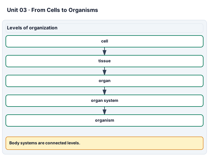
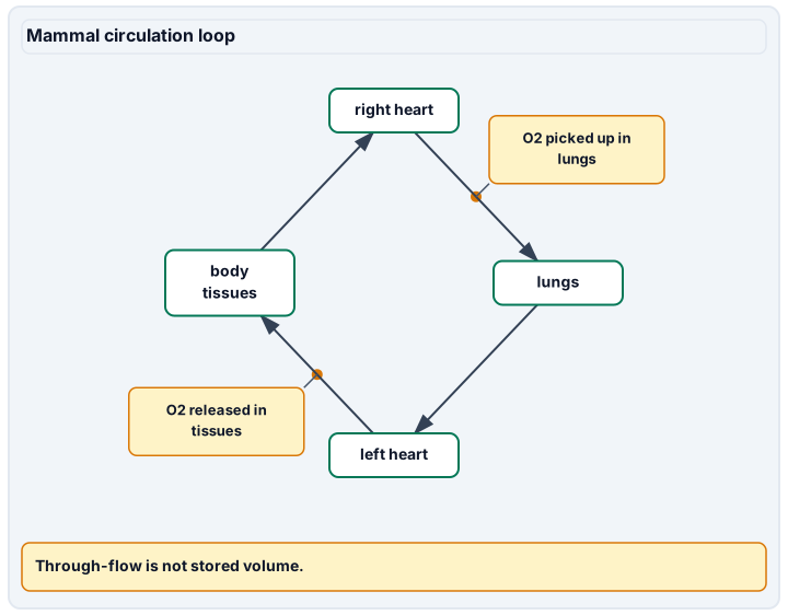
Diagram description
Cells form tissues, tissues form organs, organs form organ systems, and organ systems support an organism. Blood circulates from the right heart to lungs, left heart, body tissues, and back to the right heart. Oxygen is picked up at lungs and released at tissues. The model computes recirculating through-flow, not a distinct stored blood volume.
Data, provenance, and assumptions
| Beats counted | Counting interval (seconds) | Volume per beat (mL) | Flow duration (minutes) |
|---|---|---|---|
| 36 | 30 | 70 | 10 |
Worked model
36 beats in 30 seconds gives 72 beats/minute. With 70 mL/beat, flow is 5,040 mL/minute = 5.04 L/minute. In ten minutes, 50.4 L passes the pump. This is recirculating through-flow, not 50.4 L of distinct blood stored in the organism.
Numerical calibration
- 72 beats/minute
- 5040 mL/minute
- 50.4 L passing the modeled pump in ten minutes
Attempt the assigned level
Try the tasks before reading the calibration. These are practice answers, not a secure examination; use a new dataset or changed assumption for the assessed transfer.
Foundation: typically grades 7-8
- Build a paper flow map with right heart, lungs, left heart, body tissues, and return; label oxygen entry and carbon dioxide exit.
- Submit one cell-to-organism chain and a six-system function list; connect digestive nutrient supply and respiratory gas exchange to circulation.
Check after your attempt
- Right heart -> lungs -> left heart -> body -> right heart; gas exchange occurs across respiratory surfaces and capillaries, not by the heart making oxygen.
- Cardiac muscle cell -> cardiac muscle tissue -> heart -> circulatory system -> organism. Nutrients and gases reach cells through cooperating systems.
Core
- Calculate beats/minute, mL/minute, and litres passing in ten minutes. Mark which values are assumptions and which are derived.
- Explain how a thin exchange barrier and large surface area serve function. Contrast the total flow with the amount of blood present at one instant.
Check after your attempt
- 72 beats/minute, 5,040 mL/minute, 50.4 L in ten minutes. The constant 70 mL/beat is an input, not measured student physiology.
- Short diffusion distance and broad exchange area support transfer. Repeated circulation allows cumulative flow to exceed the stored volume.
Extension
- Recalculate ten-minute flow if stroke volume is 60 rather than 70 mL while rate stays 72 beats/minute.
- Explain why a plausible flow calculation supports recirculation but cannot by itself map every vessel or prove the cause of a person's symptoms.
Check after your attempt
- 72 x 60 x 10 / 1000 = 43.2 L, showing dependence on an assumed volume.
- The model omits pressure, variable flow, detailed exchange, and clinical measurements. Additional anatomical evidence is required; it is not a diagnostic test.
History, reading, and writing connection
Original response: Write a claim-evidence-limit paragraph comparing the opening argument in Harvey's Chapter VIII and the quantity argument in Chapter IX with the modern OpenStax circulation diagram. Cite 1628 for the work, identify the later English translation, and use the 50.4 L synthetic result to explain recirculation. State one anatomical fact supplied by the modern diagram that your calculation alone cannot establish.
Write in your own words or use an approved accessible equivalent. Cite a specific assigned section or figure, identify its evidence, and state one limitation or counterargument. Use the AI practice contract only for permitted coaching, never to invent observations or write the assessed response.
Transfer to a new case
A water pump recirculates 2 L/minute through a closed 5 L model for ten minutes. Did the model create 20 L of new water?
Calibration: No. Twenty litres passed a point, while the same water recirculated. The distinction between flow and stored quantity also matters in the circulation example.
Evidence to retain
Levels of organization: ordered example chain. Major body systems: six-function list. How systems work together: directional oxygen/nutrient flow map. Structure fits function: thin-barrier and surface-area explanation. Lab technique (body-system modeling): separately observe the student assembling and defending the paper model; supplied diagrams do not prove that performance. Retain rate calculations and assumptions, the Harvey/OpenStax comparison and transfer; integration is assessed separately.
Record units, calculations, source/date, uncertainty, and what is measured versus inferred. A simulation or supplied dataset must stay labeled as such. This is reading, paper-model, and data work, not authorization to collect pond water, swab people, culture organisms, dissect, expose wildlife, or ingest study materials. Use only instructor-approved prepared slides, images, or in-room materials for separately observed practical skills. Supplied data are not your observations.
Return to all eight learning pathways. Print this unit page for the student lessons; the linked five-page packet remains the separate assessment companion.
Unit mastery rubric
| Criterion | Developing | Proficient | Mastery |
|---|---|---|---|
| Levels of organization | Thinks the body is just one big lump of cells. | Lists cells, tissues, and organs but mixes up the order. | Puts the levels in order — cells → tissues → organs → organ systems → organism — with an example of each. |
| Major body systems | Can name only one or two body systems. | Names several systems but not what each one does. | Names the major systems — digestive, circulatory, respiratory, skeletal, muscular, nervous — and explains the main job of each. |
| How systems work together | Treats each body system as if it works alone. | Names connections but confuses flow with stored quantity. | Traces gas and nutrient transport between systems and calculates a supplied flow rate with units and assumptions. |
| Structure fits function | Doesn’t link a body part’s shape to its job. | Notices a shape but can’t explain why it helps. | Explains how thin exchange barriers and large surface area support gas transfer between lungs and blood. |
| Lab technique (body-system modeling) | Builds a model that leaves out key parts or connections. | Builds a model but can’t explain how the parts link up. | Builds a clear model of a body system and uses it to explain how the parts work together. |
| Integration (cross-domain) | Makes no supported connection between the source and the science. | Uses the source but needs help connecting evidence, writing, or limitations to the science. | Independently connects History, Reading, and Writing using a cited source, appropriate evidence, a limitation, and a scientific explanation. |
Integration is reported separately and cannot lower the science grade or block a science demonstration pass. Science and practical criteria determine that pass. Use the integration guide's evidence checklist for the separately reported criterion.
“Lung surfaces exchange gases with blood across thin barriers; the heart circulates it to tissues. In the supplied model, 36 beats in 30 seconds and 70 mL per beat give 5,040 mL per minute. The ten-minute flow of 50.4 L is recirculation, not that much distinct blood stored in the body.”
“The heart pumps blood and the lungs are for breathing. I’m not sure how they’re connected. A tissue is kind of like an organ, right?”
You demonstrate this unit by building and explaining models of the body systems — showing how cells build up to whole systems and how those systems work together — aloud, not on a multiple-choice test. A criterion counts as mastered only when you can both build the model and explain the biology it shows. Mastery is demonstrated, not awarded.
A 5-page clipboard packet — unit overview, key terms, the mastery rubric, anchor examples, and a score sheet you can print and grade against.